Review
. 2013 Nov 1;2013(3):249-60.
doi: 10.5339/gcsp.2013.32. eCollection 2013.

The science and practice of cardiopulmonary bypass: From cross circulation to ECMO and SIRS

Affiliations
Review

The science and practice of cardiopulmonary bypass: From cross circulation to ECMO and SIRS

Prakash P Punjabi et al. Glob Cardiol Sci Pract. .
No abstract available


Figures

Figure 1.
Figure 1.
Controlled cross-circulation. (A) Patient with sites of arterial and venous cannulations. (B) Donor with sites of arterial and venous cannulations. (C) Motor pump to control exchange of blood between the patient and donor. (D) Close-up of the patient's heart, showing canula to draw venous blood from both the superior and inferior venae cavae. Arterial blood from donor entered patient's body through canula in the left subclavian artery.
Figure 2.
Figure 2.
Overview of cardiopulmonary bypass circuit and its various components.
Figure 3.
Figure 3.
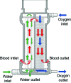
Schematic represenation of hollow fibre micro-porous oxygenator and oxygen, water, and blood flow.
Figure 4.
Figure 4.
Aspirated blood passes through a cardiotomy reservoir and micro-filter before returning to the venous reservoir.
Figure 5.
Figure 5.
Arterial and venous lines are clamped when cardiopulmonary bypass machine is not in active use.
Figure 6.
Figure 6.
Various venous cannulas tip orientations.
Figure 7.
Figure 7.
Insertion of canulae in bicaval cannulation and single atrial or Cavotrial cannulation.
Figure 8.
Figure 8.
Depiction of oxygenated and deoxygenated blood flow in ECMO cervical canulation.
Figure 9.
Figure 9.
Veno-Arterial and Veno-Venous: Extracorporeal membrane oxygenation circuit.
Figure 10.
Figure 10.
Pumpless extracorporeal lung assist with connected oxygen line.
Figure 11.
Figure 11.
Overview of various pathways to inflammatory response intiated by contact actication of blood proteins.
Figure 12.
Figure 12.
Contact activation of coagulation cascade via intrinstic coagulation pathway, proinflammatory cytokines and bleeding.
Figure 13.
Figure 13.
Edothelial cell-leukocyte adhesion cascade and its three distinctive steps.
Figure 14.
Figure 14.
Neutrophil transmigration involves movement into vascular compartment into the tissues of vital organs.
Figure 15.
Figure 15.
Use of intravital microscopy to image and assess endothelial cell interaction.
Figure 16.
Figure 16.
Sites of Aprotinin action as anti-inflammatory agent in coagulation cascade.

References

    1. Sixth National Adult Cardiac Surgical Database Report: Demonstrating Quality 2008: Bridgewater B, Keogh B, et al; July 2009 – ISBN 1-903968-23-2
    1. Jacobi C. Ein betrag zur technik der kunstlichen durchblutung uberlebender organe. Arch Exp Pathol (Leipzig) 1895;31:330–348.
    1. Brukhonenko SS, Terebinsky S. Experience avec la tete isole du chien: I. Techniques et conditions des experiences. J Physiol Pathol Genet. 1929;27:31.
    1. Lillehei CW. Historical development of cardiopulmonary bypass. Cardiopulm Bypass. 1993;1:26.
    1. Lillehei CW, Cohen M, Warden HE, Ziegler NR, Varco RL. The results of direct vision closure of ventricular septal defects in eight patients by means of controlled cross circulation. Surg Gynecol Obstet. 1955;101:446–466. - PubMed