Review
. 2013 Nov 1;2013(3):303-15.
doi: 10.5339/gcsp.2013.37. eCollection 2013.

Cardiomyocyte proliferation vs progenitor cells in myocardial regeneration: The debate continues

Affiliations
Review

Cardiomyocyte proliferation vs progenitor cells in myocardial regeneration: The debate continues

Konstantinos Malliaras et al. Glob Cardiol Sci Pract. .

Abstract

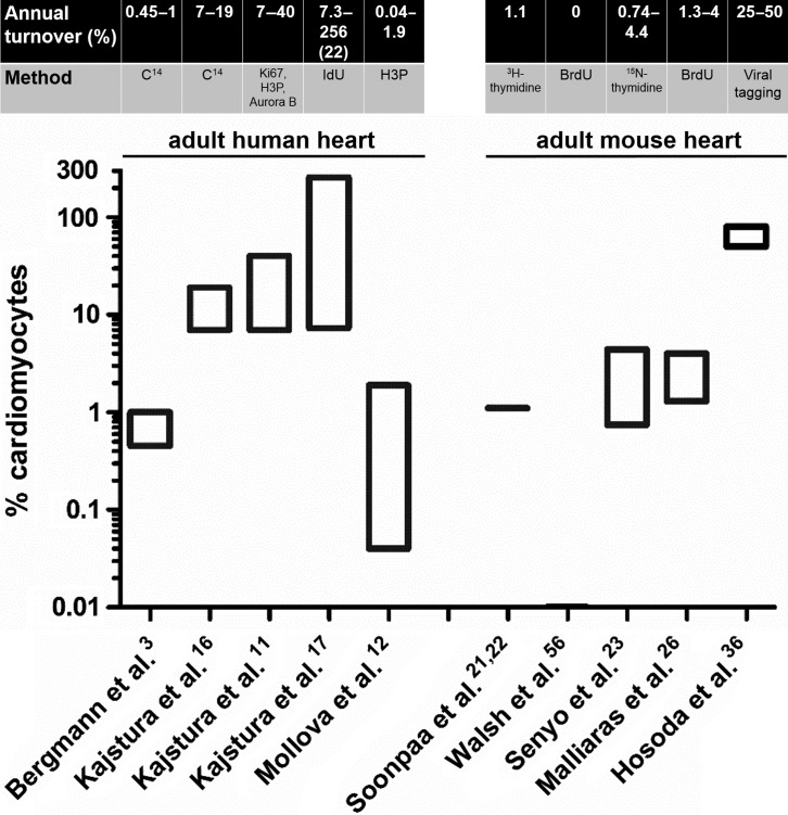
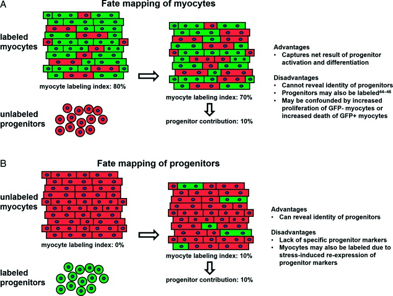
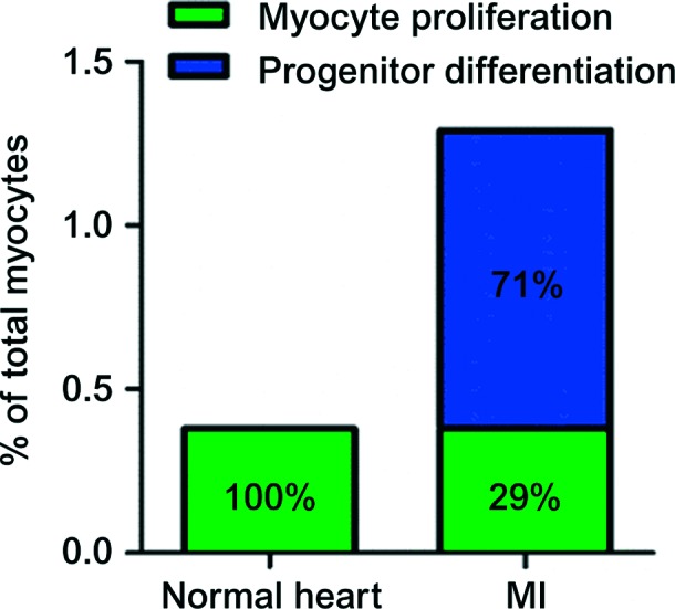
In recent years, several landmark studies have provided compelling evidence that cardiomyogenesis occurs in the adult mammalian heart. However, the rate of new cardiomyocyte formation is inadequate for complete restoration of the normal mass of myocardial tissue, should a significant myocardial injury occur, such as myocardial infarction. The cellular origin of postnatal cardiomyogenesis in mammals remains a controversial issue and two mechanisms seem to be participating, proliferation of pre-existing cardiomyocytes and myogenic differentiation of progenitor cells. We will discuss the relative importance of these two processes in different settings, such as normal ageing and post-myocardial injury, as well as the strengths and limitations of the existing experimental methodologies used in the relevant studies. Further clarification of the mechanisms underlying cardiomyogenesis in mammals will open the way for their therapeutic exploitation in the clinical field, with the scope of myocardial regeneration.


Figures

Figure 1.
Figure 1.
Annual myocyte turnover in the adult human and mouse heart. The estimated annual rates of turnover and the method of quantification are depicted. Only the turnover rates measured in adult hearts are presented. Note the logarithmic scale of the y axis. The number in parenthesis in the study by Kajstura et al. indicates the average rate of turnover measured in that study.
Figure 2.
Figure 2.
Cycling resident cardiomyocytes in the adult mouse heart. Two genetically labeled (with GFP, in green) pre-existing, α-sarcomeric actinin (αSA)-positive cardiomyocytes have incorporated BrdU in their nuclei (arrows). Z-stacks reveal that the BrdU+ nuclei belong to cardiomyocytes (αSA). The image is obtained from Malliaras et al.
Figure 3.
Figure 3.
Fate mapping strategies to study endogenous cardiac regeneration: pros and cons. A: Pre-existing cardiomyocytes are genetically labeled. Any contributions to the myocyte pool arising from cardiomyogenic differentiation of unlabeled progenitors undergo results in dilution of GFP+ myocytes by GFP −  myocytes (generated from unlabeled precursors). B: Endogenous progenitors (but not pre-existing myocytes) are genetically labeled in a prospective manner, enabling direct visualization of their future contributions to the myocyte pool.
Figure 4.
Figure 4.
Origins of postnatal cardiomyogenesis in the normal and infarcted heart. Both absolute rates (5 weeks, y axis) and relative magnitudes (percentages inside bars) are presented. In the normal mouse heart, cardiomyocyte turnover occurs through proliferation of resident cardiomyocytes. After MI, cardiomyocyte cycling increases, but the majority of new cardiomyocytes arise from recruited endogenous stem cells. The basal rate of cardiomyocyte proliferation measured in the normal heart is not included in the bar of the infarcted heart; the latter only demonstrates the additional myocytes generated post-myocardial injury. The image is modified from Malliaras et al.
Figure 5.
Figure 5.
Confounding factors when studying myocyte turnover. DNA incorporation of nucleoside analogues or nuclear expression of cell-cycle proteins, while demonstrating cell-cycle activity, does not necessarily translate into genuine cell division and proliferation; confounding factors that need to be accounted for include polyploidization (DNA proliferation without karyokinesis and cytokinesis) and bi/multinucleation (DNA proliferation and karyokinesis without cytokinesis) and to a lesser degree cell fusion and DNA repair.

References

    1. Oberpriller JO, Oberpriller JC. Response of the adult newt ventricle to injury. J Exp Zool. 1974;187:249–253. - PubMed
    1. Poss KD, Wilson LG, Keating MT. Heart regeneration in zebrafish. Science. 2002;298:2188–2190. - PubMed
    1. Bergmann O, Bhardwaj RD, Bernard S, Zdunek S, Barnabé-Heider F, Walsh S, Zupicich J, Alkass K, Buchholz BA, Druid H, Jovinge S, Frisén J. Evidence for cardiomyocyte renewal in humans. Science. 2009;324:98–102. - PMC - PubMed
    1. Porrello ER, Mahmoud AI, Simpson E, Hill JA, Richardson JA, Olson EN, Sadek HA. Transient regenerative potential of the neonatal mouse heart. Science. 2011;331:1078–1080. - PMC - PubMed
    1. Porrello ER, Mahmoud AI, Simpson E, Johnson BA, Grinsfelder D, Canseco D, Mammen PP, Rothermel BA, Olson EN, Sadek HA. Regulation of neonatal and adult mammalian heart regeneration by the miR-15 family. Proc Natl Acad Sci USA. 2013;110:187–192. - PMC - PubMed