. 2014:2014:536482.
doi: 10.1155/2014/536482. Epub 2014 Feb 9.

Flow cytometry assessment of in vitro generated CD138+ human plasma cells

Affiliations

Flow cytometry assessment of in vitro generated CD138+ human plasma cells

Rayelle Itoua Maïga et al. Biomed Res Int. 2014.

Abstract

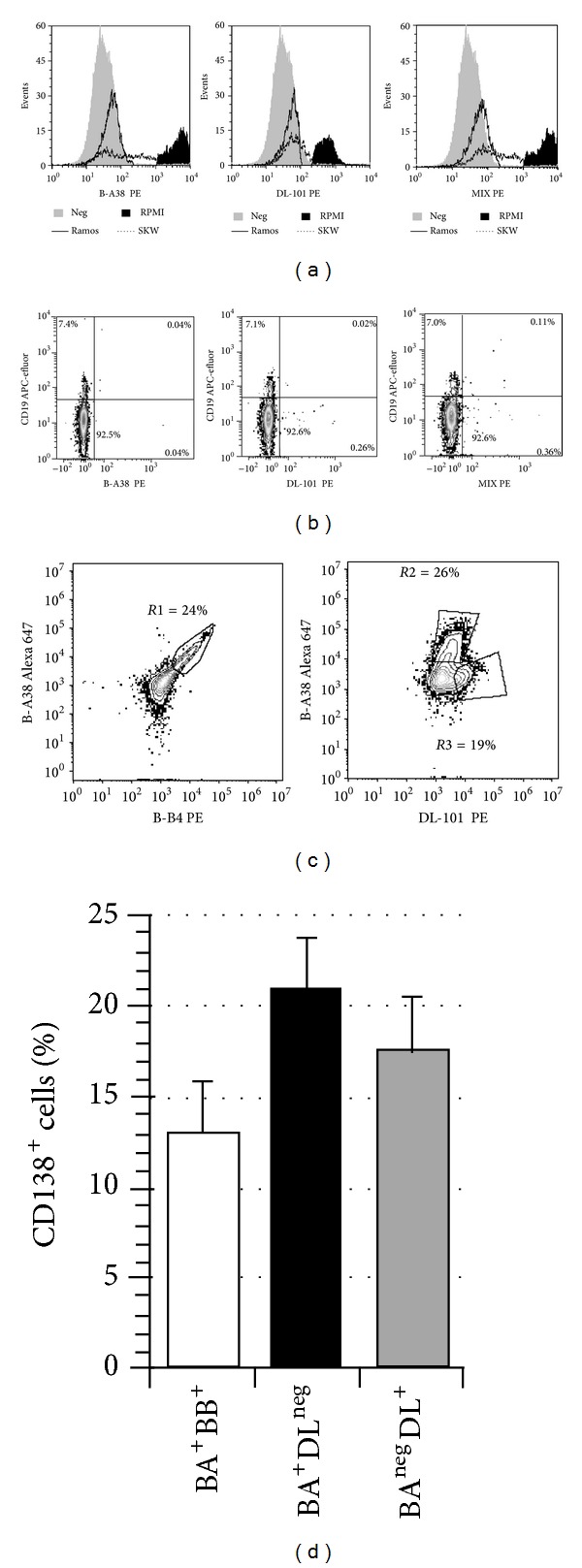
The in vitro CD40-CD154 interaction promotes human B lymphocytes differentiation into plasma cells. Currently, CD138 is the hallmark marker enabling the detection of human plasma cells, both in vitro and in vivo; its presence can be monitored by flow cytometry using a specific antibody. We have developed a culture system allowing for the differentiation of memory B lymphocytes. In order to detect the newly formed plasma cells, we have compared their staining using five anti-CD138 monoclonal antibodies (mAbs). As a reference, we also tested human cell lines, peripheral blood mononuclear cells, and bone marrow samples. The five anti-CD138 mAbs stained RPMI-8226 cells (>98%) with variable stain index (SI). The highest SI was obtained with B-A38 mAb while the lowest SI was obtained with DL-101 and 1D4 mAbs. However, the anti-CD138 mAbs were not showing equivalent CD138(+) cells frequencies within the generated plasma cells. B-A38, B-B4, and MI-15 were similar (15-25%) while DL-101 mAb stained a higher proportion of CD138-positive cells (38-42%). DL-101 and B-A38 mAbs stained similar populations in bone marrow samples but differed in their capacity to bind to CD138(high) and CD138(lo) cell lines. In conclusion, such cellular fluctuations suggest heterogeneity in human plasma cell populations and/or in CD138 molecules.


Figures

Figure 1
Figure 1
CD138+ cells staining in newly generated plasma cells. Switched memory B lymphocytes were cultured for 24 days as described in M&M. The cells were stained with one of four PE-conjugated anti-CD138 mAbs, namely, B-A38, B-B4, DL-101, and MI-15, or with a mix of B-A38 and DL-101 (MIX). The cells were fixed, kept in the dark, and analyzed 18 h to 20 h later. CD138 expression profile obtained with B-A38 for one sample out of six independent experiments is compared in (a) with B-B4 and MI-15 staining and in (b) with DL-101 and the mix of DL-101 and B-A38 (MIX). (c) The median fluorescence intensity (MFI) for each CD138 staining condition is presented as the mean ± S.E.M. for B-B4 and MI-15 staining (n = 6) and for B-A38, DL-101, and the MIX staining (n = 10) as observed for in vitro generated plasma cells. No significant differences were obtained for MFI values for any possible comparisons. (d) The CD138+ cells frequency in the viable cells (Pacific blue negative) is presented as the mean ± S.E.M for each staining condition (n = 6 or n = 10 as in panel (c)). Multiple comparisons were performed using a Bonferroni correction. Differences were observed between DL-101 and BA-38, B-B4, and MI-15 (indicated by **; P < 0.001) as well as the MIX and BA-38, B-B4, and MI-15 (indicated by *; P < 0.001).
Figure 2
Figure 2
DL-101 binding on human cells lines, blood CD19+ cells, and in vitro plasma cells. RPMI-8226, SKW6.4, and Ramos cell lines were mixed in equal proportion and stained with PE-conjugated B-A38 and DL-101 alone or combined (MIX). The staining profiles are presented for the three conditions (a). The same three anti-CD138 conditions were also used on four independent samples of blood mononuclear cells. (b) Sytox Blue, CD45, and CD19 were first used to gate the viable CD19+CD45+ cells which were then analyzed for CD138 expression. (c) Differentiated B lymphocytes, prepared as in Figure 1, were simultaneously stained with PerCP-conjugated CD19 antibodies, APC-efluor-conjugated B-A38, and either PE-conjugated B-B4 or DL-101. Analyses were done on gated CD19+ viable cells. An example representative of 5 independent experiments is shown. R1 gate included double-positive cells for B-A38 (BA) and B-B4 (BB) mAbs while R2 and R3 gates included single-positive cells for B-A38 (BA) or DL-101 (DL) mAbs, respectively. (d) The gates established in (c), namely, BA+BB+ (R1), BA+DLneg (R2), and BAnegDL+ (R3), were used to establish the proportion of these CD138+ cells subsets for five independent B lymphocyte cultures. The results are shown as the mean ± S.E.M. No significant differences were observed between all possible comparisons (Tukey-Kramer multiple comparisons test, P = 0.2220).
Figure 3
Figure 3
B-A38 and DL-101 are both present on bone marrow plasma cells. Four bone marrow samples were used to test the capacity of DL-101 to detect CD138+ cells. (a) The analysis was done on viable cells, using Sytox Blue exclusion, and then on gated CD45+CD19lo cells, which represented less than 0.5% of total cells. (b) Profiles are showed for B-A38 and DL-101 PE-conjugated mAbs for one sample gated on CD19loCD45+ region. The mean frequency ± S.E.M for CD138+ cells was 78.7% ± 1.2% for B-A38 and 59.3% ± 2.1% for DL-101. (c) Frequencies of total CD19loCD138+ are shown for all bone marrow samples according to B-A38 and DL-101; the mean ± S.E.M. was 0.20% ± 0.06% and 0.11% ± 0.05%, respectively. No significant differences were observed between the B-A38 and DL-101 staining in these four bone marrow samples (nonparametric Kruskal-Wallis test).
Figure 4
Figure 4
Titration of DL-101 and B-A38 staining on RPMI-8226. RPMI-8226 cells were stained with PE-conjugated DL-101 from eBioscience (DL-101a) and BioLegend (DL-101b) as well as B-A38. The manufacturer recommended volume was used as the reference (1, as indicated by arrow) and DL-101b and B-A38 were used at 0.25-, 0.5-, 1-, 2-, and 5-fold and DL-101a was used at 0.20-, 0.4-, 1-, 2-, and 4-fold. SI was calculated as described in Table 1.

References

    1. Carey DJ. Syndecans: multifunctional cell-surface co-receptors. Biochemical Journal. 1997;327(1):1–16. - PMC - PubMed
    1. Gattei V, Godeas C, Degan M, Rossi FM, Aldinucci D, Pinto A. Characterization of anti-CD138 monoclonal antibodies as tools for investigating the molecular polymorphism of syndecan-1 in human lymphoma cells. British Journal of Haematology. 1999;104(1):152–162. - PubMed
    1. O’Connell FP, Pinkus JL, Pinkus GS. CD138 (Syndecan-1), a plasma cell marker—immunohistochemical profile in hematopoietic and nonhematopoietic neoplasms. The American Journal of Clinical Pathology. 2004;121(2):254–263. - PubMed
    1. Wijdenes J, Vooijs WC, Clément C, et al. A plasmocyte selective monoclonal antibody (B-B4) recognizes syndecan-1. British Journal of Haematology. 1996;94(2):318–323. - PubMed
    1. Costes V, Magen V, Legouffe E, et al. The Mi15 monoclonal antibody (anti-syndecan-1) is a reliable marker for quantifying plasma cells in paraffin-embedded bone marrow biopsy specimens. Human Pathology. 1999;30(12):1405–1411. - PubMed

LinkOut - more resources