Review
. 2014:2014:819324.
doi: 10.1155/2014/819324. Epub 2014 Feb 13.

Advance of molecular imaging technology and targeted imaging agent in imaging and therapy

Affiliations
Review

Advance of molecular imaging technology and targeted imaging agent in imaging and therapy

Zhi-Yi Chen et al. Biomed Res Int. 2014.

Abstract

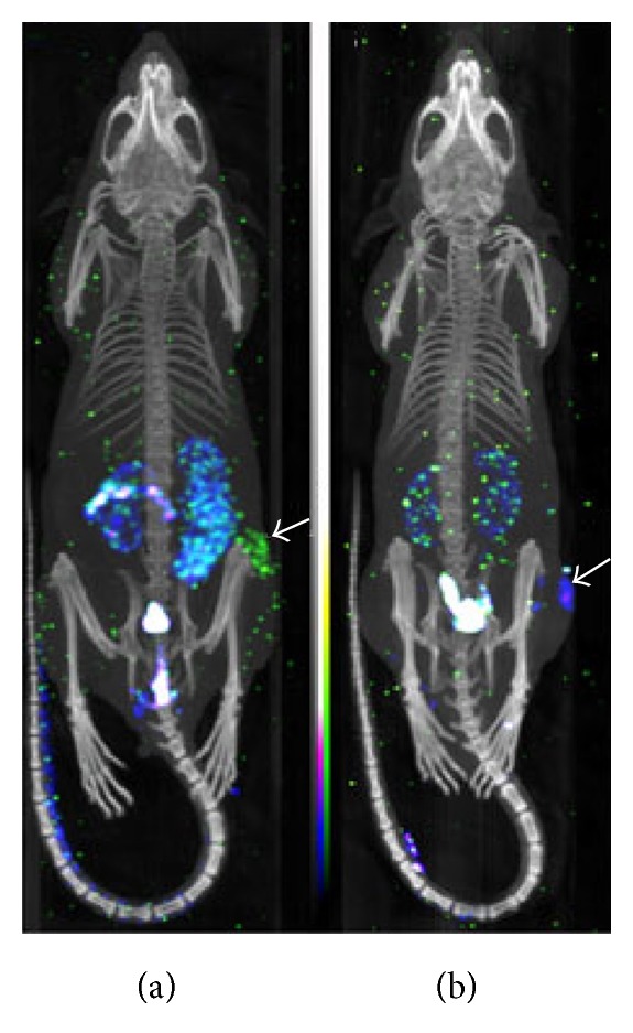
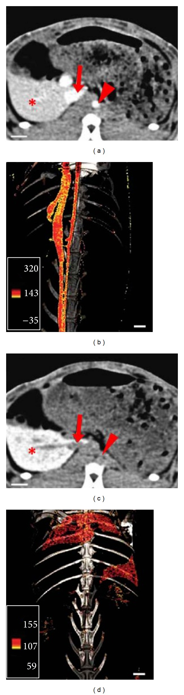
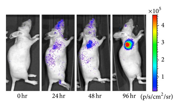
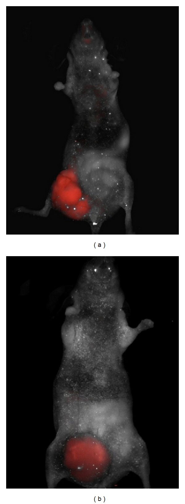
Molecular imaging is an emerging field that integrates advanced imaging technology with cellular and molecular biology. It can realize noninvasive and real time visualization, measurement of physiological or pathological process in the living organism at the cellular and molecular level, providing an effective method of information acquiring for diagnosis, therapy, and drug development and evaluating treatment of efficacy. Molecular imaging requires high resolution and high sensitive instruments and specific imaging agents that link the imaging signal with molecular event. Recently, the application of new emerging chemical technology and nanotechnology has stimulated the development of imaging agents. Nanoparticles modified with small molecule, peptide, antibody, and aptamer have been extensively applied for preclinical studies. Therapeutic drug or gene is incorporated into nanoparticles to construct multifunctional imaging agents which allow for theranostic applications. In this review, we will discuss the characteristics of molecular imaging, the novel imaging agent including targeted imaging agent and multifunctional imaging agent, as well as cite some examples of their application in molecular imaging and therapy.


Figures

Figure 1
Figure 1
Dual-isotope SPECT/CT images of tumor-bearing mice. (a) The mice were imaged 1 h after coinjection of 1.32 nmol 177Lu—DOTA—cRGDfK (yellow-green colormap) and 1.32 nmol 111In—DOTA—cRADfK (blue-purple colormap); (b) the mice were imaged 1 h after coinjection of 1.32 nmol 111In—DOTA—cRGDfK (blue-purple colormap) and 1.32 nmol 177Lu—DOTA—cRADfK (yellow-green colormap). The white arrows indicate the locations of tumor [23].
Figure 2
Figure 2
CT molecular imaging based on iodinated nanoparticles (N1177). The kinetics and distribution of iodinated nanoparticles (N1177) in the atherosclerotic rabbit model were displayed by CT imaging. (a) 5 min after intravenous injection of N1177, CT image display aorta, and vena cava (red arrow) with a noticeable signal; (b) the reconstruction of three-dimensional CT imaging; (c) 2 h after intravenous injection of N1177, a strong signal was displayed in the spleen (∗); (d) use of color scale to reconstruct three-dimensional CT angiograms of CT scan. (adopted from Hyafil et al. [33]).
Figure 3
Figure 3
After doxorubicin treatment (5 mg/kg), bioluminescence imaging was performed to observe caspase activation in 22B-pcFluc-DEVD (adopted from Niu et al. [38]).
Figure 4
Figure 4
Nude mice was injected with SW620 CRC cells then administered intravenously with integrin α v β 3 fluorescent probe after the formation of intestinal tumor. Via a fluorescent scanner, the targeted fluorescence signals for α v β 3 were observed in vivo (a). A fluorescent-labeled antibody against the VEGFR was injected intravenously, and fluorescence was detected using a multispectral in vivo imaging system (b) (adopted from Atreya et al. [39]).
Figure 5
Figure 5
31-year-old female patient with cervical cancer had an 18F FDG-PET/CT and PET/MRI examination for restaging following radiotherapy. PET/CT showed the primary tumour in the cervix behind the urine bladder and a lymph node in the pelvis, both indicated with arrows. PET/MR exhibited the same findings but with a more precise definition of the primary tumor (adopted from Kjær et al. [55]).
Figure 6
Figure 6
T1-weighted multiple slice multiple echo weighted MRI of nude mice bearing A549 tumors before and 0.5, 2, and 6 h after intraperitoneal injection. Tumors (arrows) all showed different enhancement level at 0.5 h, but in Gd-DOTA-ASON (upper line), the enhancement remained at 6 h, while Gd-DTPA (middle line) decreased obviously from 2 h (repetition time, 561 ms, echo time, 14 ms, and field of view, 4 cm); Gd-DOTA-SON (lower line) showed a similar trend to that of Gd-DOTA-ASON but with lower enhancement (adopted from Ren et al. [61]).
Figure 7
Figure 7
PET images were performed at 0.5, 2, 5, 16, and 24 h p.i. of 64Cu-NOTA-TRC105-Fab, or 64Cu-NOTA-TRC105-Fab after treatment with a 2 mg blocking dose of TRC105 before injection (adopted from Zhang et al. [66]).
Figure 8
Figure 8
Cancer-targeted multimodality imaging through MFRAS1411 conjugated nanoparticles. Tumor-bearing mice were intravenously injected with MFR-AS1411 nanoparticles (b1) and MFR-AS1411mt (b2) nanoparticles (control group). Radionuclide images were performed at 1, 6, and 24 h after injection. Scintigraphic images of tumors in mice injected with MFRAS1411 exhibited that MFR-AS1411 nanoparticles were accumulated in the tumors but MFRAS1411mt were not. Tumor growth patterns were followed using bioluminescence signals acquired from luciferase-expressing C6 cells (a1, a2). MR images of tumor-bearing mice before (c1, d1) and after (c2, d2) injection of MFR-AS1411 were obtained. Dark signal intensities at tumor sites were detected in MFR-AS1411-injected mice (arrowhead) (adopted from Hwang et al. [73]).

References

    1. Pan D, Lanza GM, Wickline SA, Caruthers SD. Nanomedicine: perspective and promises with ligand-directed molecular imaging. European Journal of Radiology. 2009;70(2):274–285. - PubMed
    1. Osborn EA, Jaffer FA. Advances in molecular imaging of atherosclerotic vascular disease. Current Opinion in Cardiology. 2008;23(6):620–628. - PMC - PubMed
    1. Weissleder R, Pittet MJ. Imaging in the era of molecular oncology. Nature. 2008;452(7187):580–589. - PMC - PubMed
    1. Atreya R, Goetz M. Molecular imaging in gastroenterology. Nature Reviews Gastroenterology & Hepatology. 2013;10(12):704–7712. - PubMed
    1. New SEP, Aikawa E. Molecular imaging insights into early inflammatory stages of arterial and aortic valve calcification. Circulation Research. 2011;108(11):1381–1391. - PMC - PubMed

Publication types