. 2014 Sep 15;76(6):438-46.
doi: 10.1016/j.biopsych.2014.02.010. Epub 2014 Feb 22.

Abnormal cortical growth in schizophrenia targets normative modules of synchronized development

Affiliations

Abnormal cortical growth in schizophrenia targets normative modules of synchronized development

Aaron F Alexander-Bloch et al. Biol Psychiatry. .

Abstract

Background: Schizophrenia is a disorder of brain connectivity and altered neurodevelopmental processes. Cross-sectional case-control studies in different age groups have suggested that deficits in cortical thickness in childhood-onset schizophrenia may normalize over time, suggesting a disorder-related difference in cortical growth trajectories.

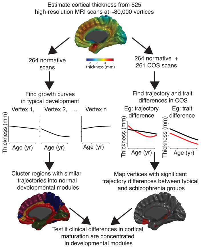
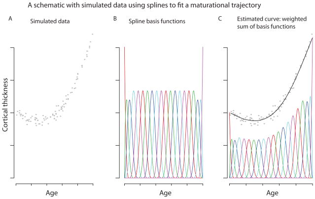
Methods: We acquired magnetic resonance imaging scans repeated over several years for each subject, in a sample of 106 patients with childhood-onset schizophrenia and 102 age-matched healthy volunteers. Using semiparametric regression, we modeled the effect of schizophrenia on the growth curve of cortical thickness in ~80,000 locations across the cortex, in the age range 8 to 30 years. In addition, we derived normative developmental modules composed of cortical regions with similar maturational trajectories for cortical thickness in typical brain development.

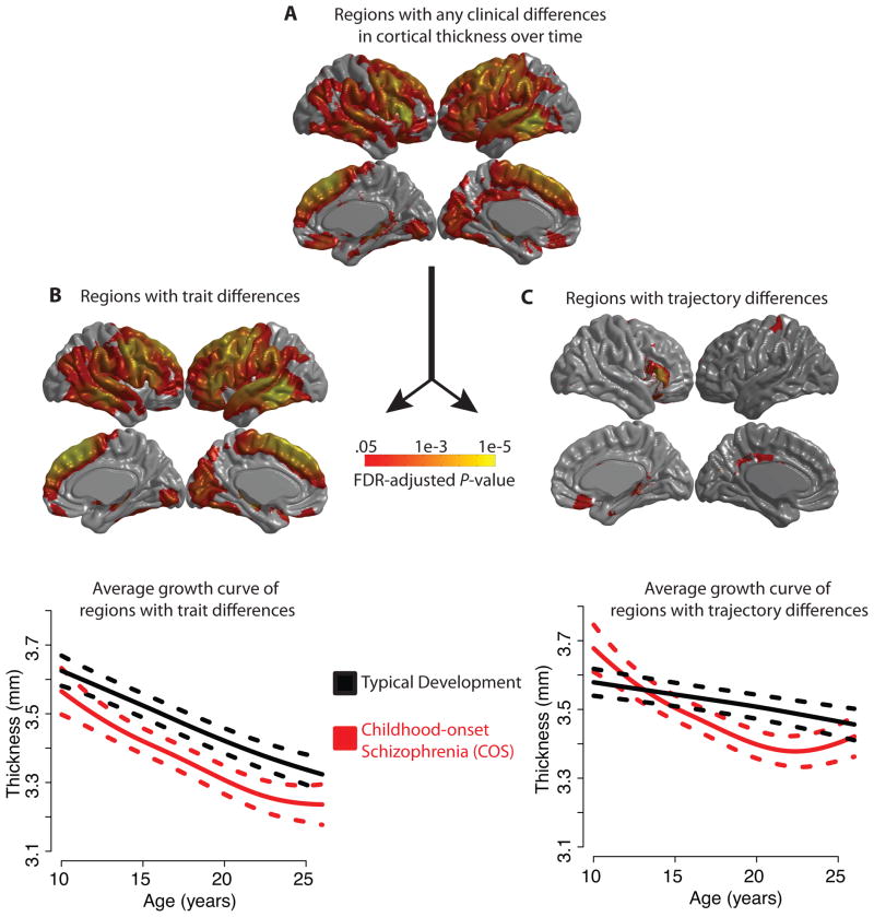
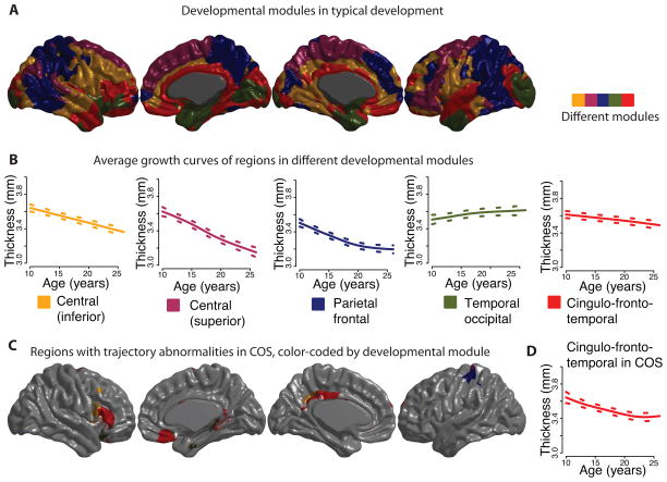
Results: We found abnormal nonlinear growth processes in prefrontal and temporal areas that have previously been implicated in schizophrenia, distinguishing for the first time between cortical areas with age-constant deficits in cortical thickness and areas whose maturational trajectories are altered in schizophrenia. In addition, we showed that when the brain is divided into five normative developmental modules, the areas with abnormal cortical growth overlap significantly only with the cingulo-fronto-temporal module.

Conclusions: These findings suggest that abnormal cortical development in schizophrenia may be modularized or constrained by the normal community structure of developmental modules of the human brain connectome.

Keywords: Neuroimaging; penalized splines; psychosis; system; topology.


Figures

Figure 1
Figure 1. Schematic of streams of analysis
As part of the intramural NIMH study of typical development and childhood-onset schizophrenia (COS), 525 high-resolution magnetic resonance imaging scans were acquired on 208 subjects, 102 with COS. For each scan, thickness was estimated at ~80,000 cortical vertices via MNI’s CIVET pipeline, and penalized splines were used to estimate maturational trajectories (thickness as a function of age). Using only healthy subjects, developmental modules were derived by clustering vertices with similarly shaped maturational trajectories. Using both healthy subjects and subjects with COS, schizophrenia-related alterations in cortical maturation were tested for all cortical vertices. Finally, it was determined whether schizophrenia-related alterations in maturational trajectories were influenced by the organization of normative developmental modules.
Figure 2
Figure 2. Spline models
A) For purposes of illustration, we used 100 simulated data points, to represent the cortical thickness of 100 subjects at different ages. B) Cubic B-spline basis functions, each in a different color. C) A weighted sum of the basis functions was used to fit a smooth curve to the simulated data.
Figure 3
Figure 3. Abnormalities of growth curves in childhood-onset schizophrenia (COS) for each of the ~80,000 cortical vertices, using penalized spline models and FDR-adjusted p-values
A) Cortical regions with any difference in the maturational trajectory in COS, either a constant trait difference or an age-varying trajectory difference. In other words, the null hypothesis H0, that βv (age) in equation (2) is identically zero, is rejected. B) Regions for which the null hypothesis H0a that βv(1)=0 in equation (3) is rejected. All of these regions are in fact thinner in COS. The plot below shows the average maturational trajectory of these regions with 95% confidence intervals. C) Regions with significant group differences in trajectory, i.e., the null hypothesis H0b that βv(2) (age) is identically zero in equation (3) is rejected. The plot below shows the average maturational trajectory of these regions with 95% confidence intervals.
Figure 4
Figure 4. Developmental modules comprised of regions with similar maturational trajectories for cortical thickness during childhood and adolescence
A k-medoids algorithm was applied to the set of estimated maturational trajectories to explore patterns of coordinated maturation across the cortex. A) As an illustration we present a map of the set of 5 developmental modules, which can be differentiated into an inferior central module (yellow), a superior central module (purple), a parietal frontal module (blue), a temporal occipital module (green), and finally a cingulo-fronto-temporal module composed of cingulate, temporal and prefrontal areas (red). B) The growth trajectory for each module is shown, averaged over all of the vertices in the module, for the typical development group of subjects. C) The cingulo-fronto-temporal module has a particularly high concentration of areas whose maturational trajectories are altered in COS (Figure 3C), although the developmental modules were calculated using only the typically developing sample. D) The growth trajectory averaged over all vertices in the cingulo-fronto-temporal module, in the COS group of subjects.

Comment in

References

    1. Haijma SV, Van Haren N, Cahn W, Koolschijn PCMP, Hulshoff Pol HE, Kahn RS. Brain Volumes in Schizophrenia: A Meta-Analysis in Over 18 000 Subjects. Schizophr Bull. 2012 doi: 10.1093/schbul/sbs118. - DOI - PMC - PubMed
    1. Glahn DC, Laird AR, Ellison-Wright I, Thelen SM, Robinson JL, Lancaster JL, et al. Meta-Analysis of Gray Matter Anomalies in Schizophrenia: Application of Anatomic Likelihood Estimation and Network Analysis. Biol Psychiatry. 2008;64:774–781. - PMC - PubMed
    1. Nenadic I, Gaser C, Sauer H. Heterogeneity of brain structural variation and the structural imaging endophenotypes in schizophrenia. Neuropsychobiology. 2012;66:44–49. - PubMed
    1. Ellison-Wright I, Bullmore E. Schizophr Res. Vol. 108. Elsevier; 2009. Meta-analysis of diffusion tensor imaging studies in schizophrenia; pp. 3–10. - PubMed
    1. Honea R, Crow TJ, Passingham D, Mackay CE. Am J Psychiatry. Vol. 162. American Psychiatric Association; 2005. Regional Deficits in Brain Volume in Schizophrenia: A Meta-Analysis of Voxel-Based Morphometry Studies; pp. 2233–2245. - PubMed

Publication types