. 2014 Apr 1;6(1):7.
doi: 10.1186/2045-824X-6-7.

Development of immortalized mouse aortic endothelial cell lines

Affiliations

Development of immortalized mouse aortic endothelial cell lines

Chih-Wen Ni et al. Vasc Cell. .

Abstract

Background: The understanding of endothelial cell biology has been facilitated by the availability of primary endothelial cell cultures from a variety of sites and species; however, the isolation and maintenance of primary mouse aortic endothelial cells (MAECs) remain a formidable challenge. Culturing MAECs is difficult as they are prone to phenotypic drift during culture. Therefore, there is a need to have a dependable in vitro culture system, wherein the primary endothelial cells retain their properties and phenotypes.

Methods: Here, we developed an effective method to prepare immortalized MAEC (iMAEC) lines. Primary MAECs, initially isolated from aortic explants, were immortalized using a retrovirus expressing polyoma middle T-antigen. Immortalized cells were then incubated with DiI-acetylated-low density lipoprotein and sorted via flow cytometry to isolate iMAECs.

Results: iMAECs expressed common markers of endothelial cells, including PECAM1, eNOS, VE-cadherin, and von Willebrand Factor. iMAECs aligned in the direction of imposed laminar shear and retained the ability to form tubes. Using this method, we have generated iMAEC lines from wild-type and various genetically modified mice such as p47phox-/-, eNOS-/-, and caveolin-1-/-.

Conclusion: In summary, generation of iMAEC lines from various genetically modified mouse lines provides an invaluable tool to study vascular biology and pathophysiology.


Figures

Figure 1
Figure 1
Scheme of mouse aortic endothelial cell isolation and immortalization.
Figure 2
Figure 2
Morphology of mouse aortic endothelial cells. (A) Aortic explants were cultured on top of collagen gel beads for 4 days. EC grew and migrated out of the aorta piece. (B) EC grown on collagen gel beads without migration seems to keep their original elongated morphology. (C-E) iMAECs collected after cell sorting were cultured for 24 h and imaged (C: phase contrast, D: DiI-Ac-LDL staining by fluorescence microscopy, and E: merged image). Scale bar = 50 μm.
Figure 3
Figure 3
Characterization of iMAEC lines by Dil-Ac-LDL staining. iMAEC lines including wild-type (iMAEC-WT), Caveolin-1 knockout (iMAEC-cav1), eNOS knockout (iMAEC-eNOS), and p47phox knockout (iMAEC-p47), were cultured. (A) Total cell lysates were collected from iMAEC lines or control cells including HUVEC, 3T3, and RASMC. Western blotting was performed using specific antibodies against Flk-1, eNOS and Cav-1. Actin serves as an internal loading control. (B) iMAECs were incubated with Dil-Ac-LDL (10 μg/mL) for 4 h, and images were taken by fluorescence microscopy. HUVEC served as positive control while 3T3 and RASMC served as negative controls. Scale bar: 50 μm. (C) Graph shows the cell shape index of HUVECs, iMAECs and primary MAECs. For cell shape index calculation, 25 cells were chosen randomly from each group and analyzed by ImageJ software. Data represent means ± standard error.
Figure 4
Figure 4
Characterization of iMAEC lines by immunostaining. iMAEC lines including iMAEC-WT, iMAEC-cav1, iMAEC-eNOS, and iMAEC-p47 were immunostained using the endothelial markers, PECAM-1, VE-Cadherin, and von Willebrand factor (vWF). Smooth muscle cell α-actin (α-SMA) was used as negative marker. HUVEC served as a positive control while 3T3 and RASMC were negative controls. Scale bar = 50 μm.
Figure 5
Figure 5
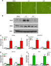
iMAEC-WT maintains shear-sensitive endothelial phenotype. iMAEC-WT were exposed to LS or OS or kept for static for 24 h. (A) iMAEC-WT aligned in the direction of the flow when exposed to LS but not OS or static control. Arrows indicate the imposed flow direction. (B) Total cell lysates were collected and protein level of KLF2 and eNOS was measured by Western blot. Data were shown as means ± standard error, n = 3. (C-F) Expression of shear-sensitive genes and microRNA under LS and OS for 24 h using early and late passages of iMAEC cells. iMAECs from early and late passages were subjected to either LS or OS for 24 h and expression of VCAM1, KLF2 and Interleukin-8(C, D, F) was determined by qPCR and normalized to 18S. (E) Graph shows the expression of shear-sensitive miRNA, miR-712 as determined by qPCR and normalized to RNU6B. Cells under static condition served as control. * p < 0.05 compared to respective LS controls.
Figure 6
Figure 6
Functional response of iMAECs to shear stress. (A) Endothelial cell tube formation; (B) endothelial cell sprouting; and (C) endothelial cell scratch wound migration assay performed using iMAECs exposed to LS or OS for 24 h. Dotted black line in B denotes the periphery of the Matrigel bead for sprouting assay. Black scale bar = 200 μm. Cells under static condition were used a controls. Data shown as means ± standard error; n = 3; * p < 0.05.
Figure 7
Figure 7
VCAM-1 expression is elevated in iMAEC-eNOS while superoxide production is diminished in iMAEC-p47. iMAEC-WT and iMAEC-p47 were stained with DHE (2 μM) for 30 min and images were acquired using fluorescence microscopy.

References

    1. Cecchi E, Giglioli C, Valente S, Lazzeri C, Gensini GF, Abbate R, Mannini L. Role of hemodynamic shear stress in cardiovascular disease. Atherosclerosis. 2011;214:249–256. doi: 10.1016/j.atherosclerosis.2010.09.008. - DOI - PubMed
    1. Dewey JCF, Gimbrone JMA, Davies PF, Bussolari SR. The Dynamic Response of Vascular Endothelial Cells to Fluid Shear Stress. J Biomech Eng. 1981;103:177–185. doi: 10.1115/1.3138276. - DOI - PubMed
    1. d'Uscio LV, Baker TA, Mantilla CB, Smith L, Weiler D, Sieck GC, Katusic ZS. Mechanism of endothelial dysfunction in apolipoprotein E-deficient mice. Arterioscler Thromb Vasc Biol. 2001;21:1017–1022. doi: 10.1161/01.ATV.21.6.1017. - DOI - PubMed
    1. Rao RM, Yang L, Garcia-Cardena G, Luscinskas FW. Endothelial-dependent mechanisms of leukocyte recruitment to the vascular wall. Circ Res. 2007;101:234–247. doi: 10.1161/CIRCRESAHA.107.151860b. - DOI - PubMed
    1. Jaffe EA, Nachman RL, Becker CG, Minick CR. Culture of human endothelial cells derived from umbilical veins. Identification by morphologic and immunologic criteria. J Clin Invest. 1973;52:2745–2756. doi: 10.1172/JCI107470. - DOI - PMC - PubMed