. 2014 Apr 1:15:7.
doi: 10.1186/1471-2091-15-7.

Functional expression and activity of the recombinant antifungal defensin PvD1r from Phaseolus vulgaris L. (common bean) seeds

Affiliations

Functional expression and activity of the recombinant antifungal defensin PvD1r from Phaseolus vulgaris L. (common bean) seeds

Erica de O Mello et al. BMC Biochem. .

Abstract

Background: Defensins are basic, cysteine-rich antimicrobial peptides that are important components of plant defense against pathogens. Previously, we isolated a defensin, PvD1, from Phaseolus vulgaris L. (common bean) seeds.

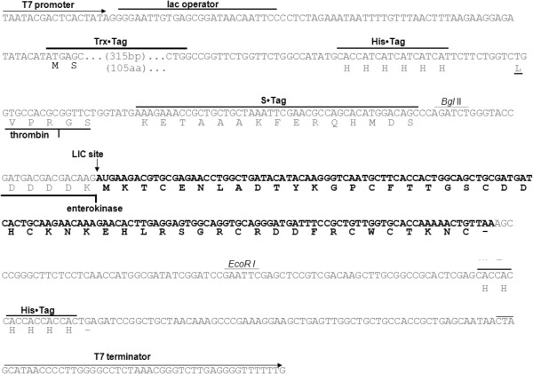
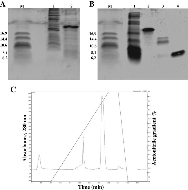
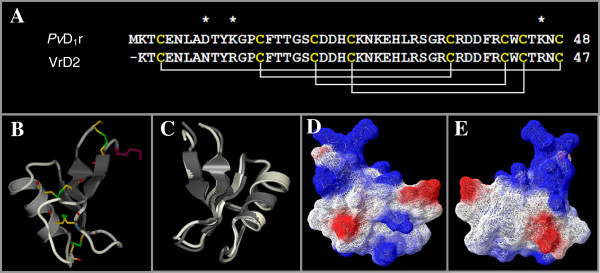
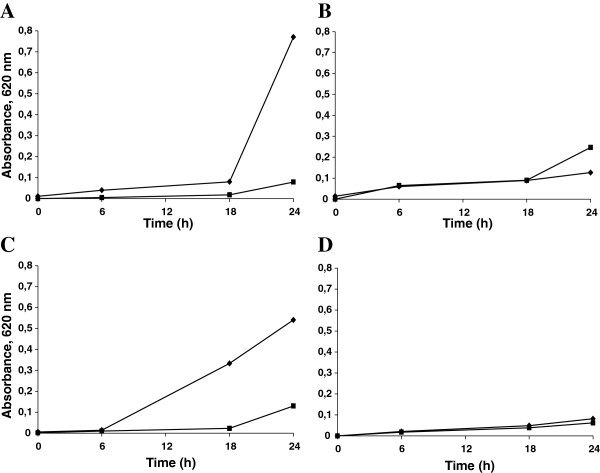
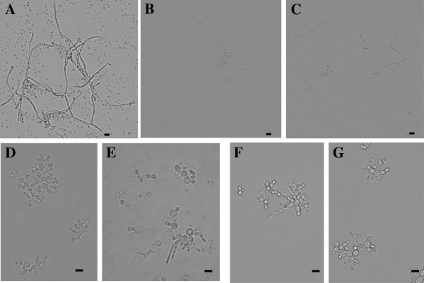
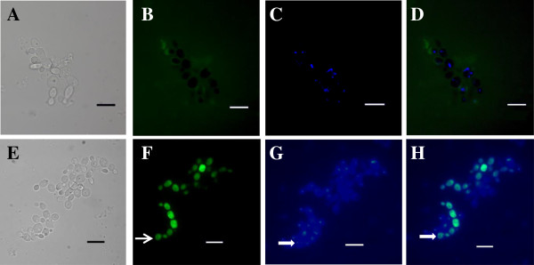
Results: The aim of this study was to overexpress PvD1 in a prokaryotic system, verify the biologic function of recombinant PvD1 (PvD1r) by comparing the antimicrobial activity of PvD1r to that of the natural defensin, PvD1, and use a mutant Candida albicans strain that lacks the gene for sphingolipid biosynthesis to unravel the target site of the PvD1r in C. albicans cells. The cDNA encoding PvD1, which was previously obtained, was cloned into the pET-32 EK/LIC vector, and the resulting construct was used to transform bacterial cells (Rosetta Gami 2 (DE3) pLysS) leading to recombinant protein expression. After expression had been induced, PvD1r was purified, cleaved with enterokinase and repurified by chromatographic steps. N-terminal amino acid sequencing showed that the overall process of the recombinant production of PvD1r, including cleavage with the enterokinase, was successful. Additionally, modeling revealed that PvD1r had a structure that was similar to the defensin isolated from plants. Purified PvD1 and PvD1r possessed inhibitory activity against the growth of the wild-type pathogenic yeast strain C. albicans. Both defensins, however, did not present inhibitory activity against the mutant strain of C. albicans. Antifungal assays with the wild-type C. albicans strains showed morphological changes upon observation by light microscopy following growth assays. PvD1r was coupled to FITC, and the subsequent treatment of wild type C. albicans with DAPI revealed that the labeled peptide was intracellularly localized. In the mutant strain, no intracellular labeling was detected.

Conclusion: Our results indicate that PvD1r retains full biological activity after recombinant production, enterokinase cleavage and purification. Additionally, our results from the antimicrobial assay, the microscopic analysis and the PvD1r-FITC labeling assays corroborate each other and lead us to suggest that the target of PvD1 in C. albicans cells is the sphingolipid glucosylceramide.


Figures

Figure 1
Figure 1
Sequence map of the cloning of PvD1 into the expression vector pET-32 EK/LIC. The coding sequence of the PvD1 (in bold) is shown linked to the LIC site of the vector pET-32 EK/LIC (indicated by a vertical arrow). The T7 promoter and terminator (horizontal arrows), lac operator, thioredoxin (Trx), six consecutive histidines (His) and (S) cleavable tags, thrombin and enterokinase protease sites, and His tag and terminator codon (-) are shown on the sequence map. The relevant amino acid sequences are shown below the nucleotide sequence. After expression and purification of the recombinant protein in this vector, all of the tags can be separated from the recombinant PvD1 by a combined treatment with enterokinase as well as another chromatographic step.
Figure 2
Figure 2
SDS-tricine gel electrophoresis analysis of PvD1r expression and purification. (A) Lane 1, protein extract of the uninduced pET-PvD1 transformed Rosetta-gami 2 bacteria; lane 2, protein extract of the induced pET-PvD1 transformed Rosetta-gami 2 bacteria. (B) Lane 1, N1 peak obtained from Ni+ affinity chromatography; lane 2, N2 peak obtained from Ni+ affinity chromatography; lane 3, N2 peak after enterokinase cleavage; lane 4, Purified PvD1r obtained from the C2C18 reversed-phase column; M - low molecular mass marker (kDa). (C) Chromatogram of the last step of the purification of PvD1r after cleavage of N2 with enterokinase. The oblique line indicates the acetonitrile gradient. The retention time of PvD1r was previously determined by purified PvD1r in Ni+-NTA agarose. The same retention time was collected and this sample presented only one band by tricine gel electrophoresis (Figure 2B4). The peak and the corresponding band are indicated by asterisks.
Figure 3
Figure 3
Modeling. (A) Alignment of the amino acid sequences of PvD1r and VrD2, a defensin isolated from Vigna radiata. The lines below the cysteines of VrD2 indicate the disulfide bound formation and paring among the cysteine residues. The three different amino acid residues between the two sequences are indicated by asterisks. (B) Three-dimensional structure of PvD1r, modeled with the Modeller program and based on the structure of the V. radiate defensin VrD2 (pdb CODE 2GL1). Dark gray represents the β-sheets; gray represents the α-helix; and light gray lines represent the unstructured elements. A methionine residue (purple) was added as a requirement for cloning proteins into the pET-32 EK/LIC vector. The four disulfide bridges are shown by the interconnection of the cysteines residues shown in yellow. The sulfur is shown in green, the nitrogen in blue and the oxygen in red. (C) Overlap of the three-dimensional structures of PvD1r (light gray) and Vigna radiata defensin 2 (VrD2) (code pdb 2GL1; in dark gray. (D and E) Three-dimensional structure of PvD1r with a surface charge; the negative surface charge is shown in red, and the positive surface charge is shown in blue. In E the structure is rotated 180º in relation to D. For an additional explanation of the color usage in this figure, please refer to the web version of the article.
Figure 4
Figure 4
Effects of PvD1 and PvD1r on the growth of the wild-type and mutant strains of Candida albicans. The absorbance at 620 nm was taken as a measure of C. albicans growth. (A)C. albicans wild-type strain; (B)C. albicans mutant strain; (♦) Control; (■) 100 μg.mL-1 of PvD1; (C)C. albicans wild-type strain; (D)C. albicans mutant strain; (♦) Control; (■) 100 μg.mL-1 of PvD1r. All of the experiments were performed in triplicate, and the standard errors were omitted for clarity (coefficients of variation were less than 20%).
Figure 5
Figure 5
Candida albicans cells, wild-type and mutant strains, visualized by light microscopy after the growth inhibition assay. (A) Wild-type control cells; (B) Wild-type cells grown in the presence of PvD1; (C) Wild-type cells grown in the presence of PvD1r; (D) Mutant control cells; (E) Mutant cells grown in the presence of PvD1; (F and G) Mutant cells grown in the presence of PvD1r. Bars = 10 μm.
Figure 6
Figure 6
Fluorescence microscopy of Candida albicans cells. (A-D) Mutant cells; (E-H) Wild-type cells. Cells were incubated for 24 h with 50 μg.ml -1 FITC-conjugated PvD1r (green fluorescence) (B and F). After the incubation period, the nuclei were stained with DAPI (blue fluorescence) (C and G). Light microscopy (A and E). Overlap of the DAPI and FITC images (D and H). The open arrow shows the FITC labeling (F); the filled arrows show the DAPI nuclei labeling (G) and the colocalization of FITC and DAPI (H). Bars = 10 μm.

References

    1. Terras FRG, Eggermont K, Kovaleva V, Raikhel NV, Osborn RW, Kester A, Rees SB, Torrekens S, Leuven FV, Vanderleyden J, Cammue BPA, Broekaert WF. Small cysteine-rich antifungal proteins from radish: their role in host defense. Plant Cell. 1995;7:573–588. doi: 10.1105/tpc.7.5.573. - DOI - PMC - PubMed
    1. Fant F, Vranken W, Broekaert W, Borremans F. Determination of the three-dimensional solution structure of Raphanus sativus antifungal protein 1 by 1H NMR. J Mol Biol. 1998;279:257–270. doi: 10.1006/jmbi.1998.1767. - DOI - PubMed
    1. Thomma BP, Cammue BPA, Thevissen K. Plant defensins. Planta. 2002;216:193–202. doi: 10.1007/s00425-002-0902-6. - DOI - PubMed
    1. Cornet B, Bonmatin J-M, Hetru C, Hoffmann JA, Ptak M, Vovelle F. Refined three dimensional solution structure of insect defensin A. Structure. 1995;3:435–448. doi: 10.1016/S0969-2126(01)00177-0. - DOI - PubMed
    1. Bontems F, Roumestand C, Boyot P, Gilquin B, Doljansky Y, Menez A, Toma F. Three-dimensional structure of natural charydotoxin in aqueus solution by 1H NMR. Charybdotoxin possesses a structural motif founf in other scorpion. Eur J Biochem. 1991;196:19–28. doi: 10.1111/j.1432-1033.1991.tb15780.x. - DOI - PubMed

Publication types

MeSH terms