. 2014 Apr 1:8:9.
doi: 10.1186/1754-1611-8-9. eCollection 2014.

Optimization of 3-D organotypic primary colonic cultures for organ-on-chip applications

Affiliations

Optimization of 3-D organotypic primary colonic cultures for organ-on-chip applications

Asad A Ahmad et al. J Biol Eng. .

Abstract

Background: New advances enable long-term organotypic culture of colonic epithelial stem cells that develop into structures known as colonoids. Colonoids represent a primary tissue source acting as a potential starting material for development of an in vitro model of the colon. Key features of colonic crypt isolation and subsequent colonoid culture have not been systematically optimized compromising efficiency and reproducibility. Here murine crypt isolation yield and quality are optimized, and colonoid culture efficiency measured in microfabricated culture devices.

Results: An optimal incubation time of 60 min in a chelating buffer released 280,000 ± 28,000 crypts from the stroma of a single colon with 79.3% remaining intact. Mechanical agitation using an average acceleration of 1.5 × g liberated the highest quality crypts with 86% possessing well-defined lumens. Culture in 50% Matrigel resulted in the highest colonoid formation efficiency of 33 ± 5%. Immunostaining demonstrated that colonoids isolated under these conditions possessed stem/progenitor cells and differentiated cell lineages. Microfabrication substrates (glass, polystyrene, PDMS, and epoxy photoresists: SU-8 and 1002-F) were tested for compatibility with colonoid culture. PDMS promoted formation of 3-D colonoids containing stem/progenitor cells, while other substrates promoted outgrowth of a 2-D epithelial monolayer composed of differentiated cells.

Conclusion: Improved crypt isolation and 3-D colonoid culture, along with an understanding of colonic epithelial cell behavior in the presence of microfabrication substrates will support development of 'organ-on-a-chip' approaches for studies using primary colonic epithelium.


Figures
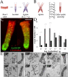
Figure 1
Figure 1
Isolation of crypts from a mouse colon. (A) Schematic of crypt isolation. The resected colon was incubated in chelating buffer, washed and then mechanically agitated. (B) Schematic of the strategy to identify the optimal acceleration intensity needed to retrieve crypts: the tissue was incubated in chelating buffer, rinsed and then sequentially agitated at different acceleration intensities. After each agitation, the crypt-rich supernatant was collected and assayed. (C) Fresh crypts isolated using an acceleration intensity of 1.5 × g displaying eGFP fluorescence, indicating Sox9 expression (green). Scale bar = 40 μm. (D) Quantification of the number of intact and broken crypts at each of the six acceleration intensities tested. Grey bars indicate intact-crypt yield and black bars indicate broken-crypt yield. (E) Brightfield images of isolated crypts isolated at different accelerations intensities. Crypts isolated using an acceleration intensity of 1.5 × g display a visible lumen, indicating unperturbed crypt morphology. Scale bar = 150 μm.
Figure 2
Figure 2
Effect of Matrigel concentration on in vitro expansion of colonic crypts into 3-D colonoids. (A) Serial overlaid brightfield and eGFP fluorescence images of the same colonoid over 1 week in culture. Scale bar = 50 μm. (B) Effect of Matrigel concentration on the percentage of crypts growing into colonoids over 1 week of culture. Squares, triangles, circles and diamonds represent 25%, 50%, 75% and 100% Matrigel, respectively. 50% Matrigel provides the optimum 3-D growth environment for the colonoids. (C) Quantification of the cell properties in the colonoids. Shown is the colonoid volume (left y axis, black) staining positive for Muc-2, ChgA or EdU divided by that positive for Hoechst 33342 when colonoids were cultured in 100% (filled bars) or 50% (open bars) Matrigel. The volume of the colonoid expressing eGFP relative to that expressing dsRed is shown on the right y-axis (grey) for colonoids cultured in 100% (filled bars) or 50% (open bars) Matrigel. (D-E) Colonoids were cultured for 1 week and then stained by immunohistochemistry for: (D) mucin-2 (goblet cell marker: green) and (E) chromogranin-A (enteroendocrine marker: green). (F) A crypt obtained from a Sox9eGFP-CAGDsRed mouse was cultured for 1 week and then imaged for eGFP fluorescence. (G) Fluorescence image of a colonoid (1 week culture) after an 8-hour EdU pulse (red). Hoescht 33442 was used as a nuclear stain (blue) in panels C-G. Scale bar = 75 μm.
Figure 3
Figure 3
Crypt-substrate interaction. (A) Representative time-lapse images of monolayer formation on the experimental substrates from crypts isolated from a Sox9eGFP-CAGDsRed mouse. eGFP and DsRed fluorescence was overlaid on brightfield microscopy images. Upon adherence to glass, oxidized polystyrene and epoxy photoresist, crypt-cells rapidly differentiate. (B) Quantification of the percentage of crypts forming a monolayer when crypts were cultured on the microfabrication substrates over 1 week. (C, D) Whole-mount immunohistochemical staining of a monolayer after 1 week in culture. Fluorescence images are shown for: mucin-2 (green, C) and chromogranin-A (green, D). (E) Fluorescence image after an 8-hour EdU pulse (red). Hoescht 33442 was used as a nuclear stain (blue) in panels C-E. Scale bars = 50 μm.

References

    1. Barker N, van de Wetering M, Clevers H. The intestinal stem cell. Genes Dev. 2008;8:1856–1864. doi: 10.1101/gad.1674008. - DOI - PMC - PubMed
    1. Garrison AP, Helmrath MA, Dekaney CM. Intestinal stem cells. J Pediatr Gastroenterol Nutr. 2009;8:2–7. doi: 10.1097/MPG.0b013e3181ad3021. - DOI - PubMed
    1. Ramalingam S, Daughtridge GW, Johnston MJ, Gracz AD, Magness ST. Distinct levels of Sox9 expression mark colon epithelial stem cells that form colonoids in culture. Am J Physiol Gastrointest Liver Physiol. 2012;8:G10–G20. doi: 10.1152/ajpgi.00277.2011. - DOI - PMC - PubMed
    1. Humphries A, Wright NA. Colonic crypt organization and tumorigenesis. Nat Rev Cancer. 2008;8:415–424. doi: 10.1038/nrc2392. - DOI - PubMed
    1. Medema JP, Vermeulen L. Microenvironmental regulation of stem cells in intestinal homeostasis and cancer. Nature. 2011;8:318–326. doi: 10.1038/nature10212. - DOI - PubMed