Genomic profile analysis of diffuse-type gastric cancers

Yeon-Su Lee et al. Genome Biol. .

Abstract

Background: Stomach cancer is the third deadliest among all cancers worldwide. Although incidence of the intestinal-type gastric cancer has decreased, the incidence of diffuse-type is still increasing and its progression is notoriously aggressive. There is insufficient information on genome variations of diffuse-type gastric cancer because its cells are usually mixed with normal cells, and this low cellularity has made it difficult to analyze the genome.

Results: We analyze whole genomes and corresponding exomes of diffuse-type gastric cancer, using matched tumor and normal samples from 14 diffuse-type and five intestinal-type gastric cancer patients. Somatic variations found in the diffuse-type gastric cancer are compared to those of the intestinal-type and to previously reported variants. We determine the average exonic somatic mutation rate of the two types. We find associated candidate driver genes, and identify seven novel somatic mutations in CDH1, which is a well-known gastric cancer-associated gene. Three-dimensional structure analysis of the mutated E-cadherin protein suggests that these new somatic mutations could cause significant functional perturbations of critical calcium-binding sites in the EC1-2 junction. Chromosomal instability analysis shows that the MDM2 gene is amplified. After thorough structural analysis, a novel fusion gene TSC2-RNF216 is identified, which may simultaneously disrupt tumor-suppressive pathways and activate tumorigenesis.

Conclusions: We report the genomic profile of diffuse-type gastric cancers including new somatic variations, a novel fusion gene, and amplification and deletion of certain chromosomal regions that contain oncogenes and tumor suppressors.


Figures

Figure 1
Figure 1
Whole genome distribution of somatic mutations and duplication or deletion events in diffuse-type gastric cancers (DGCs). All the somatic mutations, including duplication/deletion events, which were found in the 14 DGC genomes, are merged in the circus plot. From outside to inside, the plot presents the following characteristics: chromosome ideograms, frequency of cumulative amplification or deletion events (black, amplification; red, deletion), and the number of somatic non-synonymous single nucleotide variations (nsSNVs), indels, and SNVs in splice sites for each gene. Black triangles indicate highly mutated genes. Orange triangles denote oncogenes, and blue triangles indicate the tumor suppressors.
Figure 2
Figure 2
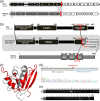
TSC2-RNF216 fusion gene breakage. (a) Exon structure of the TSC2-RNF216 fusion gene. The numbers in the boxes are the exon numbers of each gene. Red lines indicate the fusion points. (b) Protein domain structure of the TSC2-RNF216 fusion protein. The Rap-GAP domain of TSC2 was broken, and RNF216 had a frameshift mutation causing premature termination by the interchromosomal rearrangement. (c) Structure of the TSC2 Rap-GAP domain. The red region is the remaining Rap-GAP domain region, and the gray region is the Rap-GAP domain that is deleted in the TSC2-RNF216 fusion gene. (d) RNA sequence of the TSC2-RNF216 fusion gene. Position 136 is shown as N. Either an A or G base produces a termination codon (TAA or TAG). (e) Verification of the TSC2-RNF216 fusion transcript in RNA (cDNA) by means of PCR amplification and electrophoresis.
Figure 3
Figure 3
The duplication region of the MDM2 gene on chromosome 12 in samples D-01 T and D-02 T. (a) Mapping depth plots of the two chromosomes. (b) Thin black spikes were read at mapping depth of 2000-base width. The y-axis shows relative depth. Each unit represents approximately 30 times sequencing depth. (c) Gene positions and names around the amplified regions. The black bands show gene locations. (d)MDM2 transcript levels in tumor and adjacent normal tissue paired samples and normal cell lines. Quantitative RT-PCR was used to measure MDM2 mRNA levels in samples D-01 and D-02 (containing amplified MDM2 regions), D-04, D-05, D-10, I-03, and I-04 (without amplified MDM2 regions), and three normal cell lines (HDF, HMEC, and Hs 738.St/Int). Error bars were calculated from two separated experiments of triplicate reactions.
Figure 4
Figure 4
Structure of the CDH1 protein and EC1-2 junction. (a) The full-length E-cadherin protein has 882 amino acid residues in 7 domains. Sites of non-synonymous mutations and deletions are shown with red lines. (b) Red lines and triangles indicate non-synonymous mutations in extracellular cadherin (EC) domains. (c) CDH1 has five EC domains (EC1–EC5, which form a β-barrel structure) and four EC junctions (EC1-2, EC2-3, EC3-4, and EC4-5). The green spheres represent Ca2+ ions. The red and blue spheres represent somatic mutations found in this study and previously reported mutations found in hereditary diffuse-type gastric cancer, respectively. (d, e) CDH1 mutation sites in the EC1-2 junction. In the case of the D221G mutation, oxygen atoms of the aspartic acid side chain, which normally interact with Ca2+, are absent when the aspartic acid residue is replaced with a glycine. In the case of the D257N mutation, the two oxygen atoms of the Asp side chains become one oxygen atom and one nitrogen atom when aspartic acid is replaced with asparagine. In the N256S mutation, the oxygen atom of the asparagine side chain is preserved, but the distance between the oxygen atom and the Ca2+ ion is increased from 2.52 Å to 3.73 Å. All structures were drawn by using PyMOL Molecular Graphics System (v0.99rc6; Schrödinger LLC).

References

    1. Ferlay J, Soerjomataram I, Ervik M, Dikshit R, Eser S, Mathers C, Rebelo M, Parkin DM, Forman D, Bray F. GLOBOCAN 2012: Estimated Cancer Incidence, Mortality and Prevalence Worldwide in 2012. http://globocan.iarc.fr. - PubMed
    1. Lauren P. The Two histological main types of gastric carcinoma: diffuse and so-called intestinal-type carcinoma. An attempt at a histo-clinical classification. Acta Pathol Microbiol Scand. 1965;64:31–49. - PubMed
    1. Crew KD, Neugut AI. Epidemiology of gastric cancer. World J Gastroenterol. 2006;12:354–362. - PMC - PubMed
    1. Parkin DM, Bray F, Ferlay J, Pisani P. Estimating the world cancer burden: Globocan 2000. Int J Cancer. 2001;94:153–156. doi: 10.1002/ijc.1440. - DOI - PubMed
    1. Ushijima T, Sasako M. Focus on gastric cancer. Cancer Cell. 2004;5:121–125. doi: 10.1016/S1535-6108(04)00033-9. - DOI - PubMed

Publication types

MeSH terms