. 2014 Aug;39(9):2252-62.
doi: 10.1038/npp.2014.76. Epub 2014 Apr 1.

Opposing effects of acute versus chronic blockade of frontal cortex somatostatin-positive inhibitory neurons on behavioral emotionality in mice

Affiliations

Opposing effects of acute versus chronic blockade of frontal cortex somatostatin-positive inhibitory neurons on behavioral emotionality in mice

Amelie Soumier et al. Neuropsychopharmacology. 2014 Aug.

Abstract

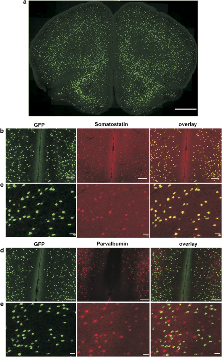
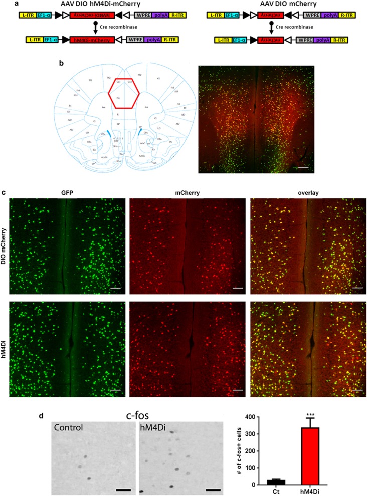
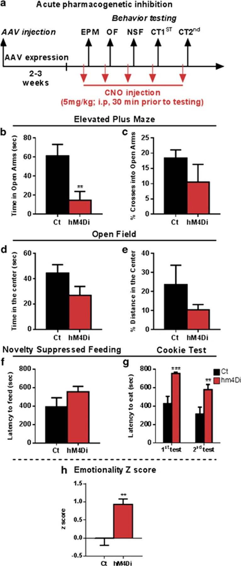
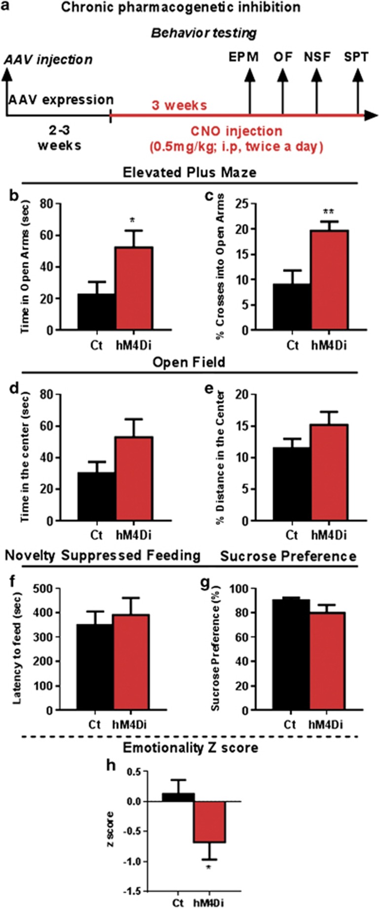
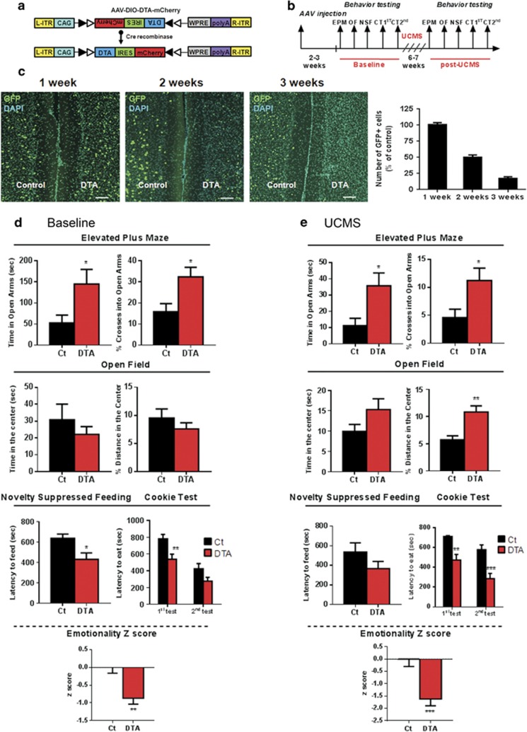
Reduced expression of somatostatin (SST) is reported across chronic brain conditions including major depression and normal aging. SST is a signaling neuropeptide and marker of gamma-amino butyric acid (GABA) neurons, which specifically inhibit pyramidal neuron dendrites. Studies in auditory cortex suggest that chronic reduction in dendritic inhibition induces compensatory homeostatic adaptations that oppose the effects of acute inhibition. Whether such mechanisms occur in frontal cortex (FC) and affect behavioral outcome is not known. Here, we used two complementary viral vector strategies to examine the effects of acute vs chronic inhibition of SST-positive neurons on behavioral emotionality in adult mice. SST-IRES-Cre mice were injected in FC (prelimbic/precingulate) with CRE-dependent adeno-associated viral (AAV) vector encoding the engineered Gi/o-coupled human muscarinic M4 designer receptor exclusively activated by a designer drug (DREADD-hM4Di) or a control reporter (AAV-DIO-mCherry) for acute or chronic cellular inhibition. A separate cohort was injected with CRE-dependent AAV vectors expressing diphtheria toxin (DTA) to selectively ablate FC SST neurons. Mice were assessed for anxiety- and depressive-like behaviors (defined as emotionality). Results indicate that acute inhibition of FC SST neurons increased behavioral emotionality, whereas chronic inhibition decreased behavioral emotionality. Furthermore, ablation of FC SST neurons also decreased behavioral emotionality under baseline condition and after chronic stress. Together, our results reveal opposite effects of acute and chronic inhibition of FC SST neurons on behavioral emotionality and suggest the recruitment of homeostatic plasticity mechanisms that have implications for understanding the neurobiology of chronic brain conditions affecting dendritic-targeting inhibitory neurons.


Figures

Figure 1
Figure 1
SST-cre:Ai6 mice allows conditional manipulation of somatostatin (SST) expressing neurons. (a) GFP expression in a frontal cortex (FC) coronal section from an adult SST-ires-cre mice crossed with a Zsgreen cre-dependent reporter mice. Scale bar: 1 mm. (b, c) Immunostaining of GFP (green) and SST (red) shows colocalization in cortical neurons (overlay, yellow). (d, e) Immunostaining of GFP (green) and parvalbumin (red) in GFP-expressing neurons (overlay) reveals non-overlapping expression. Scale bars: 1 mm in (a), 100 μm in (b, d) and 20 μm in (c, e). A full color version of this figure is available at the Neuropsychopharmacology journal online.
Figure 2
Figure 2
Cortical SST cell targeting by DREADD-hm4Di. Panel (a) represents the design of hM4Di-mCherry AAV (left) and mCherry AAV (right) vectors employing the DIO strategy. Two pairs of heterotypic, antiparallel loxP recombination sites achieve cre-mediated transgenes inversion and expression under the control of EF1α promoter. L-ITR: left-inverted terminal repeat, EF1α: Human elongation factor-1 alpha, WPRE: woodchuck hepatitis virus post-transcriptional regulatory element, R-ITR: right-inverted terminal repeat. (b) Schematic coronal section illustrating the injection site (left) of the imaged area in the FC. mCherry fluorescence was detected in the cingulate and prelimbic areas following bilateral injection into SST-cre:Ai6 mice (right). Scale bar: 500 μm. (c) mCherry (top) and hM4Di receptors (bottom) were selectively/exclusively expressed in cortical SST neurons as illustrated by GFP (green) and mCherry (red) co-expression (overlay, yellow). Scale bar: 100 μm. (d) Representative c-fos immunoreactivity from SST:cre;Ai6 mice with mCherry (control) or hm4Di AAV vectors, 90 min following CNO injection (5 mg/kg, i.p.). Scale bars: 40 μm. Activation of hm4Di receptors by CNO increased the number of c-fos+ cells (***p<0.0001, n=6/8 mice per group). Data are mean±SEM. A full color version of this figure is available at the Neuropsychopharmacology Journal online.
Figure 3
Figure 3
Acute pharmacogenetic inhibition of SST neurons increases anxiety-like behavior and overall emotionality. (a) Schematic of the experimental design used to study the acute effect of SST cells inhibition on behavior. All mice were CNO treated (5 mg/kg, i.p.) 30 min before behavioral testing. Behavioral results associated with acute pharmacogenetic inhibition of SST cells on elevated plus maze (time spent in open arms in b, percent (%) of crosses into open arms in c), open field (time spent in the center in d, % of distance traveled in the center in e), latency to eat in the novelty-suppressed feeding (f) and cookie test (g). (h) Resulting integrated emotionality z-score (**p<0.01, ***p<0.0001, n=9/10 mice per group; data are mean±SEM). A full color version of this figure is available at the Neuropsychopharmacology journal online.
Figure 4
Figure 4
Chronic pharmacogenetic inhibition of SST neurons reduces anxiety-like behavior and overall emotionality. (a) Schematic of the experimental design used to study the chronic effect of SST cells inhibition on behavior. Mice received a dose of CNO (0.5 mg/kg, i.p., twice a day, 8 h apart). Behavioral results associated with chronic pharmacogenetic inhibition of SST cells on elevated plus maze (time spent in open arms in b and % of crosses into open arms in c), open field (time spent in the center in d and % of distance traveled in the center in e), latency to eat in the novelty-suppressed feeding (f) and cookie test (g). (h) Resulting integrated emotionality z score (*p<0.05, **p<0.01, n=8 mice per group; data are mean±SEM). A full color version of this figure is available at the Neuropsychopharmacology journal online.
Figure 5
Figure 5
Selective ablation of cortical SST neurons reduces anxiety-like behavior and overall emotionality under baseline and chronic stress conditions. (a) Design of DTA-mCherry AAV employing the DIO strategy, which uses two pairs of heterotypic, antiparallel loxP recombination sites to achieve cre-mediated transgenes inversion and expression under the control of CAG promoter. L-ITR: left-inverted terminal repeat, CAG: CMV early enhancer/chicken beta actin, WPRE: woodchuck hepatitis virus posttranscriptional regulatory element, R-ITR: right-inverted terminal repeat. (b) Schematic of the experimental design used to study the effect of SST cells suppression on behavior. Mice were tested twice: before (baseline) and after unpredictable chronic stress paradigm (UCMS). (c) Illustration of GFP (green) and DAPI (blue) immunostaining in the vehicle-control side and following 1, 2, and 3 weeks of DTA injection. The quantification of GFP cells 1, 2, and 3 weeks after DTA injection expressed as the percent of control (n=3 per condition) is shown to the right. Scale bar: 100 μm. Behavioral results associated with genetic ablation of SST cells under baseline condition (d) and after UCMS (e). Note that the longitudinal aspects of the study (pre- and post-stress in same cohorts), rather than cross-sectional, preclude direct comparison of pre- vs post-stress effects, as variations in baseline behavior 6–7 weeks apart frequently occur and could be misconstrued here as ‘stress effect' if presented together. Instead the contrasts of interest ‘DTA vs control treatments' are presented under baseline and stress conditions. (*p<0.05, **p<0.01, ***p<0.001 n=13/14 mice per group; data are mean±SEM). A full color version of this figure is available at the Neuropsychopharmacology journal online.

References

    1. Alexander GM, Rogan SC, Abbas AI, Armbruster BN, Pei Y, Allen JA, et al. Remote control of neuronal activity in transgenic mice expressing evolved G protein-coupled receptors. Neuron. 2009;63:27–39. - PMC - PubMed
    1. Armbruster BN, Li X, Pausch MH, Herlitze S, Roth BL. Evolving the lock to fit the key to create a family of G protein-coupled receptors potently activated by an inert ligand. Proc Natl Acad Sci USA. 2007;104:5163–5168. - PMC - PubMed
    1. Atasoy D, Betley JN, Su HH, Sternson SM. Deconstruction of a neural circuit for hunger. Nature. 2012;488:172–177. - PMC - PubMed
    1. Bajbouj M, Lisanby SH, Lang UE, Danker-Hopfe H, Heuser I, Neu P. Evidence for impaired cortical inhibition in patients with unipolar major depression. Biol Psychiatry. 2006;59:395–400. - PubMed
    1. Brockschnieder D, Lappe-Siefke C, Goebbels S, Boesl MR, Nave KA, Riethmacher D. Cell depletion due to diphtheria toxin fragment A after Cre-mediated recombination. Mol Cell Biol. 2004;24:7636–7642. - PMC - PubMed

Publication types

MeSH terms