

doi: 10.1364/ol.39.001473.
Urogenital photoacoustic endoscope
Affiliations
- PMID: 24690816
- PMCID: PMC4009352
- DOI: 10.1364/ol.39.001473
Item in Clipboard
Urogenital photoacoustic endoscope
Opt Lett.
.
Abstract
Photoacoustic (PA) endoscopy for human urogenital imaging has the potential to diagnose many important diseases, such as endometrial and prostate cancers. We have specifically developed a 12.7 mm diameter, rigid, side-scanning PA endoscopic probe for such applications. The key features of this endoscope are the streamlined structure for smooth cavity introduction and the proximal actuation mechanism for fast scanning. Here we describe the probe's composition and scanning mechanism and present in vivo experimental results suggesting its potential for comprehensive clinical applications.
Figures
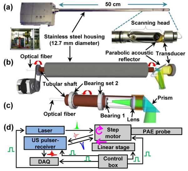
Photo (a) and schematic (b) of the PA endoscopic probe developed for human urogenital system imaging (Media 1). (c) Schematic of the rotary junction (Bearing 1) and light guiding optics. (d) Block diagram showing the peripheral systems.
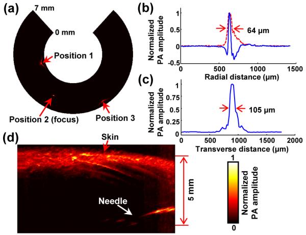
(a) PA B-scan image of the 20 μm thick tungsten wire imaged at three locations. (b) Typical PA A-line signal (blue) and its Hilbert-transformed signal (i.e., radial LSF) for the tungsten wire target located at the focal point (Position 2). (c) Corresponding transverse LSF of the tungsten wire. (d) PA image of a black needle inserted into the inner leg of a rat.
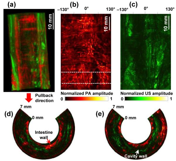
(a) Coregistered PA and US volumetric image from a rabbit colorectum acquired in vivo over a 53 mm range with a 27 mm image diameter (Media 2). The lower portion of the image corresponds to the anus. (b, c) PA- and US-RMAP images of (a). (d, e) Representative combined PA and US B-scan images near the location indicated by the dashed line in (b).
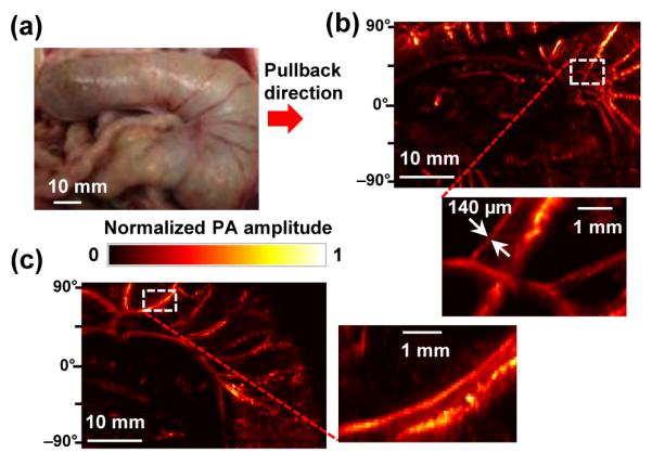
(a) Photo of the rat large intestine imaged ex vivo. (b, c) PA-RMAP images of (a). All the PA signals in the presented
References
-
- Cannistra SA, Niloff JM. Cancer of the uterine cervix. N. Engl. J. Med. 1996;334(no. 16):1030–1038. - PubMed
Publication types
MeSH terms
Grants and funding
LinkOut - more resources
Full Text Sources
Other Literature Sources
