Case Reports
. 2014 Oct;134(10):2570-2578.
doi: 10.1038/jid.2014.164. Epub 2014 Apr 1.

Epithelial inflammation resulting from an inherited loss-of-function mutation in EGFR

Affiliations
Case Reports

Epithelial inflammation resulting from an inherited loss-of-function mutation in EGFR

Patrick Campbell et al. J Invest Dermatol. 2014 Oct.

Abstract

Epidermal growth factor receptor (EGFR) signaling is fundamentally important for tissue homeostasis through EGFR/ligand interactions that stimulate numerous signal transduction pathways. Aberrant EGFR signaling has been reported in inflammatory and malignant diseases, but thus far no primary inherited defects in EGFR have been recorded. Using whole-exome sequencing, we identified a homozygous loss-of-function missense mutation in EGFR (c.1283 G>A; p.Gly428Asp) in a male infant with lifelong inflammation affecting the skin, bowel, and lungs. During the first year of life, his skin showed erosions, dry scale, and alopecia. Subsequently, there were numerous papules and pustules--similar to the rash seen in patients receiving EGFR inhibitor drugs. Skin biopsy demonstrated an altered cellular distribution of EGFR in the epidermis with reduced cell membrane labeling, and in vitro analysis of the mutant receptor revealed abrogated EGFR phosphorylation and EGF-stimulated downstream signaling. Microarray analysis on the patient's skin highlighted disturbed differentiation/premature terminal differentiation of keratinocytes and upregulation of several inflammatory/innate immune response networks. The boy died at the age of 2.5 years from extensive skin and chest infections as well as electrolyte imbalance. This case highlights the major mechanism of epithelial dysfunction following EGFR signaling ablation and illustrates the broader impact of EGFR inhibition on other tissues.


Figures
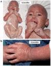
Figure 1
Figure 1. Clinical features of the patient demonstrating inflammation, erosions, papules and pustules
(a) At 12 months of age the infant has extensive erosions and markedly reduced scalp hair and eyebrows. He is also receiving total parenteral nutrition. (b) At 22 months there is a confluent papular eruption, particularly on the limbs, with numerous pustules. Consent to publish these photographs was obtained from the infant’s mother.
Figure 2
Figure 2. The homozygous mutation p.Gly428Asp results in acanthosis, intra-epidermal edema, and loss of keratinocyte cell membrane labeling for EGFR
(a) Semithin section of patient skin from the thigh reveals acanthosis and hyperkeratosis compared to age and site-matched control skin (scale bars = 50 μm). (b) Ultrastructurally, there is widening of spaces between adjacent keratinocytes in the lower epidermis (asterisks; scale bar = 2 μm). (c) Immunostaining for EGFR in patient epidermis shows loss of keratinocyte membrane staining compared to control skin (scale bars = 50 μm). (d) Sanger sequencing reveals a homozygous missense mutation in EGFR. (e) Schematic representation of the functional domains and encoding exons and the site of the pathogenic mutation in this patient. TM = transmembranous domain.
Figure 3
Figure 3. The mutation in EGFR renders the receptor unstable and susceptible to endocytosis
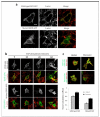
(a) Confocal microscopy was performed using GFP-tagged constructs of wild-type or mutant EGFR (green) and F-actin (red) in MCF-7 cells under normal growth conditions. (b) Images were then taken following stimulation of the cells with EGF at specified time points to assess the localization of EGFR within the cell cytoplasm or at the cell membrane. (c) Confocal microscope images were also taken for wild-type and mutant EGFR-GFP constructs (green) in cells stained for surface EGFR (red) in DMSO (dimethyl sulfoxide) control or Dynasore-treated cells (to inhibit endocytosis). The colocalization between EGFR antibody staining and either wild-type or mutant EGFR-GFP constructs was then quantified in DMSO control or Dynasore-treated cells using Pearson’s correlation coefficient (Panel D lower). * p<0.001 vs WT-EGFR; scale bars = 10μm.
Figure 4
Figure 4. The mutation in EGFR reduces signal transduction and cell proliferation
(a) Western blotting was performed for p-EGFR, EGFR, p-ERK, ERK, p-Akt and Akt on lysates from untransfected (U), wild-type EGFR (WT) or mutant EGFR (MUT) transfected MCF-7 cells after EGF stimulation for the indicated times. MCF-7 cells express very low endogenous EGFR and therefore EGFR is undetectable in UT cells. Arrows indicate phospho and total EGFR species in top and second row blots respectively. Note the higher molecular weight species of EGFR in the WT samples in the second row blot are phosphorylated receptor and directly correlate with phosphorylated EGFR as detected in the top row blot. (b) Cell proliferation was quantified in CHO-K1 cells transfected with GFP, wild-type EGFR-GFP or mutant EGFR-GFP in starved (−), normal growth (10% fetal calf serum) or EGF-stimulated (EGF) conditions. (c) GFP-positive cells were counted and normalized against total cell number, and the cell growth rate was then calculated from these data.

Comment in

References

    1. Avraham R, Yarden Y. Feedback regulation of EGFR signalling: Decision making by early and delayed loops. Nat Rev Mol Cell Biol. 2011;12:104–17. - PubMed
    1. Adzhubei IA, Schmidt S, Peshkin L, et al. A method and server for predicting damaging missense mutations. Nat Methods. 2010;7:248–9. - PMC - PubMed
    1. Blaydon DC, Biancheri P, Di WL, et al. Inflammatory skin and bowel disease linked to ADAM17 deletion. N Engl J Med. 2011;365:1502–8. - PubMed
    1. Blobel CP. ADAMs: Key components in EGFR signalling and development. Nat Rev Mol Cell Biol. 2005;6:32–43. - PubMed
    1. Chavanas S, Bodener C, Rochat A, et al. Mutations in SPINK5, encoding a serine protease inhibitor, cause Netherton syndrome. Nat Genet. 2000;25:141–2. - PubMed

Publication types

MeSH terms

Substances