. 2014 Apr 1;9(4):e93056.
doi: 10.1371/journal.pone.0093056. eCollection 2014.

C-phycocyanin confers protection against oxalate-mediated oxidative stress and mitochondrial dysfunctions in MDCK cells

Affiliations

C-phycocyanin confers protection against oxalate-mediated oxidative stress and mitochondrial dysfunctions in MDCK cells

Shukkur M Farooq et al. PLoS One. .

Erratum in

  • PLoS One. 2014;9(7):e103361. Asokan, Devarajan [corrected to Devarajan, Asokan]

Abstract

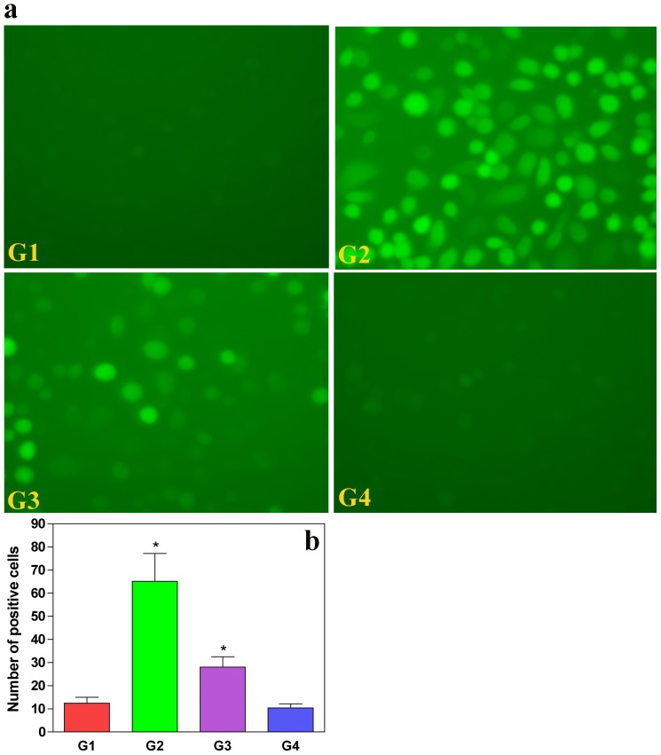
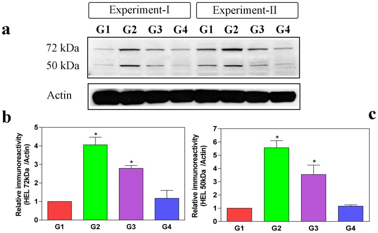
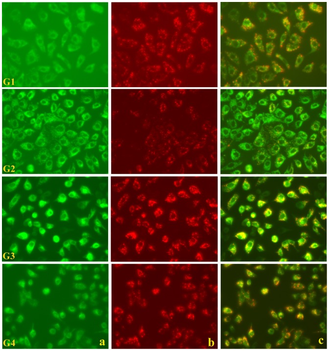
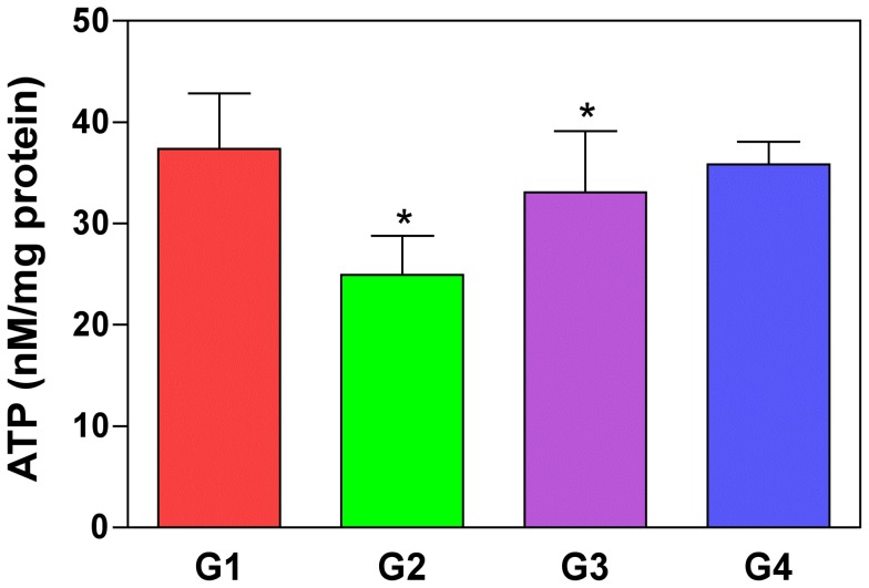
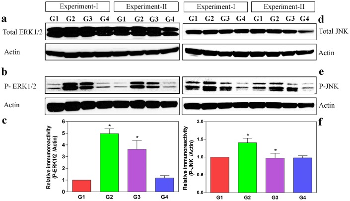
Oxalate toxicity is mediated through generation of reactive oxygen species (ROS) via a process that is partly dependent on mitochondrial dysfunction. Here, we investigated whether C-phycocyanin (CP) could protect against oxidative stress-mediated intracellular damage triggered by oxalate in MDCK cells. DCFDA, a fluorescence-based probe and hexanoyl-lysine adduct (HEL), an oxidative stress marker were used to investigate the effect of CP on oxalate-induced ROS production and membrane lipid peroxidation (LPO). The role of CP against oxalate-induced oxidative stress was studied by the evaluation of mitochondrial membrane potential by JC1 fluorescein staining, quantification of ATP synthesis and stress-induced MAP kinases (JNK/SAPK and ERK1/2). Our results revealed that oxalate-induced cells show markedly increased ROS levels and HEL protein expression that were significantly decreased following pre-treatment with CP. Further, JC1 staining showed that CP pre-treatment conferred significant protection from mitochondrial membrane permeability and increased ATP production in CP-treated cells than oxalate-alone-treated cells. In addition, CP treated cells significantly decreased the expression of phosphorylated JNK/SAPK and ERK1/2 as compared to oxalate-alone-treated cells. We concluded that CP could be used as a potential free radical-scavenging therapeutic strategy against oxidative stress-associated diseases including urolithiasis.


Conflict of interest statement

Competing Interests: The authors have declared that no competing interests exist.

Figures

Figure 1
Figure 1. Concentrations of C-phycocyanin in the presence of oxalate in MDCK cells.
Different concentrations of CP (5, 10, 20, 50 mM) in the presence of oxalate (0.1 mM) in MDCK cell line showing the effective dosage of CP at 20 mM concentration at 24 and 48 hours.
Figure 2
Figure 2. C-phycocyanin quenches oxalate-induced ROS production in MDCK cells.
(A) MDCK cells treated with oxalate were observed for ROS generation with a non-fluorescent dye DCFDA, which converts to a fluorescent DCF compound during oxidative stress (panel G2). This is compared with panel G1 (normal conditions). Panel G3 shows reduced fluorescence due to reduced generation of ROS upon CP pre-treatment, which resembles normal cells. Panel G4 confirms that CP alone by itself is not cytotoxic. (B) Quantitative estimation of DCF fluorescence, oxalate-induced MDCK cells (G2) shows exaggerated fluorescence as compared to control, and reduced florescence was observed in the CP pre-treated cells (G3). Values are expressed as mean±S.D. P<0.05 were considered significant. *G2 compared with G1; G3 compared with G2. G1: Control; G2: Oxalate; G3: Oxalate+CP; G4: CP alone.
Figure 3
Figure 3. C-phycocyanin protects cells from free radical-mediated lipid peroxidation (LPO) induced by oxalate.
To test the extent of free radical-mediated cellular damage, some oxidative stress markers have been developed and recognized suggestive of lipid peroxidation (LPO); for instance, HEL (hexanoyl-lysine adduct) is one the typical aldehyde products used as a marker of LPO. Thus, LPO was assessed by Western blotting for HEL in the lysates of MDCK cells from control (G1), oxalate alone (G2), oxalate+CP (G3) and CP alone (G4). Band intensity of HEL is prominent in oxalate-alone-treated cells (G2) at 72 kDa and 50 kDa compared to control MDCK cells (G1), whereas oxalate+CP cells (G3) showed decreased HEL band intensity relative to oxalate-treated cells (G2). The densitometric analyses were performed using a ImageJ (NIH) software. Densitometry data of 72 kDa and 50 kDa were normalized using internal control actin. Values represent mean±SD derived from immunoblots of all the groups from 3 experiments. The P values for HEL 72 kDa and 50 kDa of all the groups were as follows: HEL 72 kDa: G1 vs G2 (*P = 0.017) and G2 vs G3 (*P = 0.05); HEL 50 kDa: G1 vs G2 (*P = 0.013) and G2 vs G3 (*P = 0.009). P<0.05 were considered significant (*). G1: Control; G2: Oxalate; G3: Oxalate+CP; G4: CP alone.
Figure 4
Figure 4. C-phycocyanin protects cellular mitochondrial membrane potential from oxalate-induced ROS.
Integrity of mitochondrial membrane potential in MDCK cells investigated by a mitochondrial potential-sensitive dye, JC-1, showing normal stippled spots of bright red fluorescence (G1a–c). The numbers (viz. a–c) in the panels represents the red, green and merged channel fields observed respectively. Oxalate-exposed MDCK cells observed later (G2a–c) shows the absence of characteristic red fluorescence. Cells pre-treated with CP and oxalate showing the re-appearance of red fluorescence spots following normalization of mitochondrial potential (G3a–c). This confirms that CP could protect against mitochondrial membrane oxalate induced-ROS and does not itself affect the mitochondrial potential. Cells treated with CP alone show a normal picture (G4a–c). G1: Control; G2: Oxalate; G3: Oxalate+CP; G4: CP alone.
Figure 5
Figure 5. Oxalate-induced ATP depletion restored by C-phycocyanin pre-treatment in cells.
ATP levels in cultured MDCK cells were significantly lowered when incubated with oxalate alone (G2) than control group (G1) (P = 0.02). ATP depletion was significantly restored when cells were pre-treated with CP under oxalate-induced conditions (G3) (P = 0.012). *G2 compared with G1; G3 compared with G2. P<0.05 were considered significant. G1: Control; G2: Oxalate; G3: Oxalate+CP; G4: CP alone.
Figure 6
Figure 6. C-phycocyanin inhibits the activation of JNK and ERK1/2 in oxalate-induced cells.
Activation of ERK1/2 and JNK in (MDCK cells) western blots of control (G1), oxalate-induced conditions (G2), oxalate+CP (G3) and CP alone (G4). The lysate RAB fractions were probed with antibodies for total and activated (phosphorylated) ERK1/2 and JNK. MDCK cells in all conditions had similar levels of total JNK and ERK1/2 (a & d). However, the levels of activated (phosphorylated) ERK1/2 and JNK were significantly lowered in oxalate+CP (G3) cell lysates than the oxalate alone (G2) groups (b & e). Scanning densitometry data of phosphorylated form of ERK1/2 and JNK were normalized using internal control actin. Phosphorylated ERK1/2 and JNK levels were significantly increased following oxalate-alone induction (G2), which was significantly reduced on pre-treatment with CP (G3) (c & f). However, the total ERK1/2 and JNK levels did not differ significantly in any of the groups investigated (Histogram data not shown). The Densitometric analyses were performed using a Image J NIH software. Values represent mean±SD derived from immunobots of all the groups from 4 experiments. The P values for activated (phosphorylated) ERK1/2 and JNK of all the groups were as follows: phosphorylated ERK1/2: G1 vs G2 (*P = 0.0025) and G2 vs G3 (*P = 0.027); phosphorylated JNK: G1 vs G2 (*P = 0.0089) and G2 vs G3 (*P = 0.015). P<0.05 were considered significant (*). G1: Control; G2: Oxalate; G3: Oxalate+CP; G4: CP alone.

References

    1. Sakhaee K, Maalouf NM, Sinnott B (2012) Clinical review. Kidney stones 2012: pathogenesis, diagnosis, and management. J Clin Endocrinol Metab 97: 1847–1860. - PMC - PubMed
    1. Brzica H, Breljak D, Burckhardt BC, Burckhardt G, Sabolic I (2013) Oxalate: from the environment to kidney stones. Arh Hig Rada Toksikol 64: 609–630. - PubMed
    1. Mulay SR, Kulkarni OP, Rupanagudi KV, Migliorini A, Darisipudi MN, et al. (2013) Calcium oxalate crystals induce renal inflammation by NLRP3-mediated IL-1β secretion. Journal Clin Invest 123: 236–246. - PMC - PubMed
    1. Johri N, Cooper B, Robertson W, Choong S, Rickards D, et al. (2010) An update and practical guide to renal stone management. Nephron Clin Pract 116: c159–171. - PubMed
    1. Thamilselvan V, Menon M, Thamilselvan S (2014) Oxalate at physiological urine concentrations induces oxidative injury in renal epithelial cells: Effect of α-tocopherol and ascorbic acid. BJU Int doi: 10.1111/bju.12642 - DOI - PubMed

MeSH terms