. 2014 May;124(5):1987-99.
doi: 10.1172/JCI70324. Epub 2014 Apr 1.

Transporters MCT8 and OATP1C1 maintain murine brain thyroid hormone homeostasis

Transporters MCT8 and OATP1C1 maintain murine brain thyroid hormone homeostasis

Steffen Mayerl et al. J Clin Invest. 2014 May.

Abstract

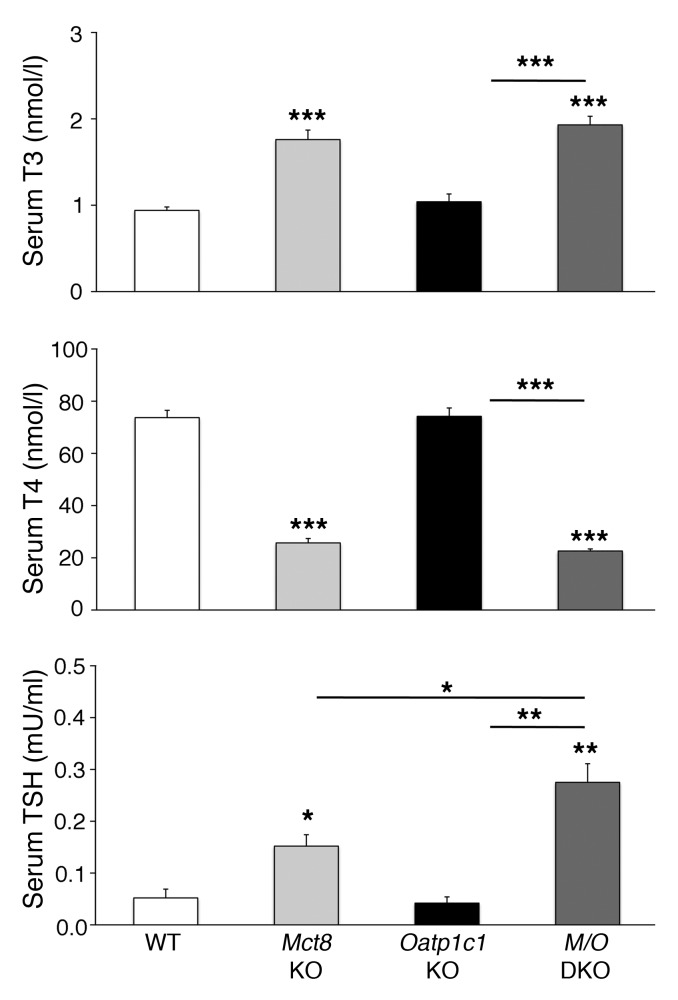
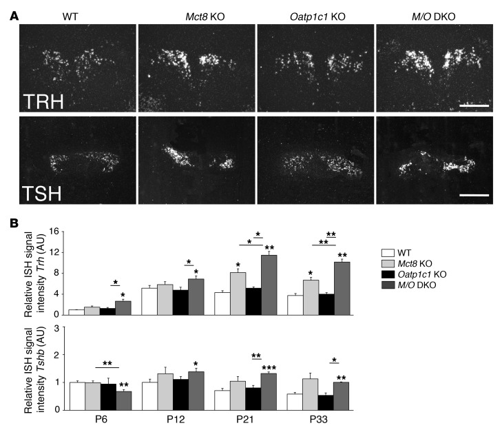
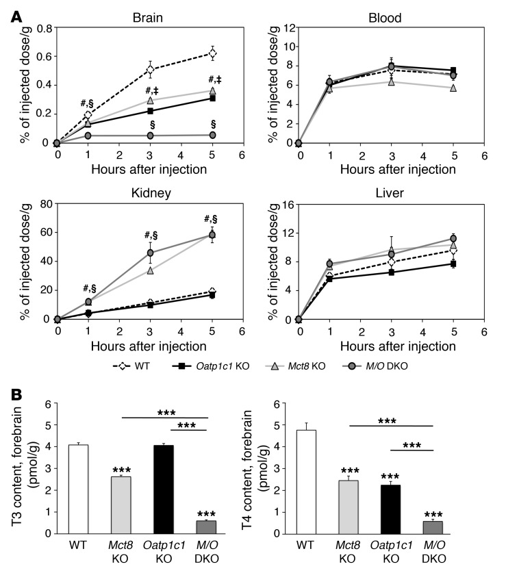
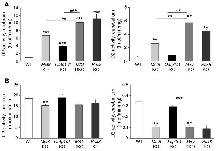
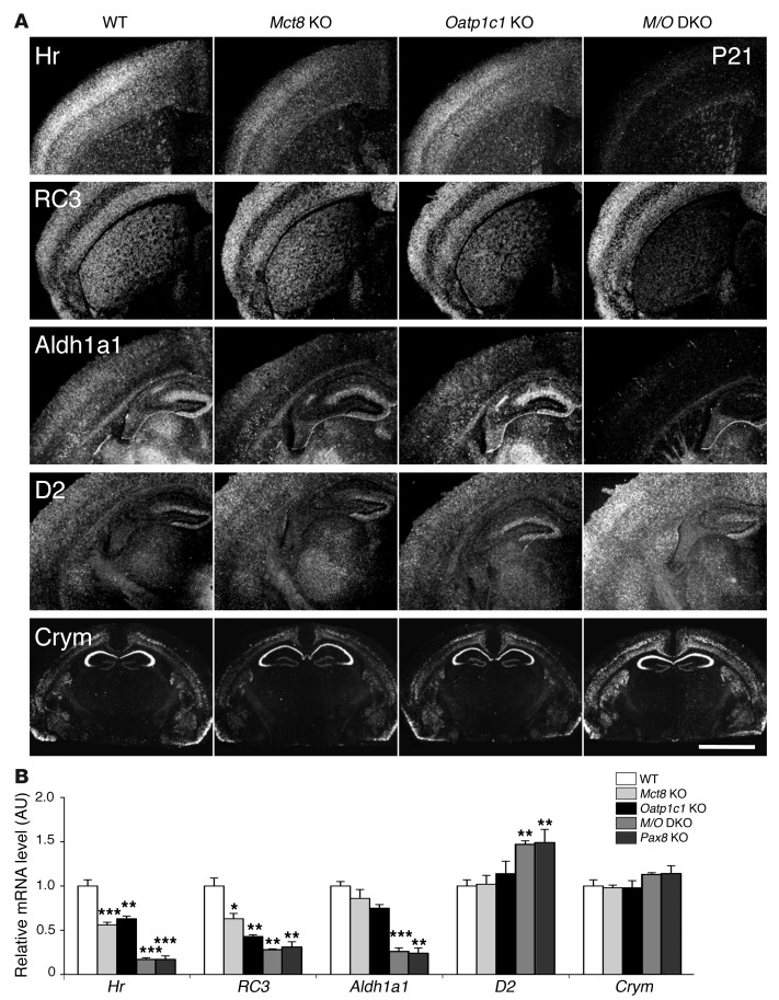
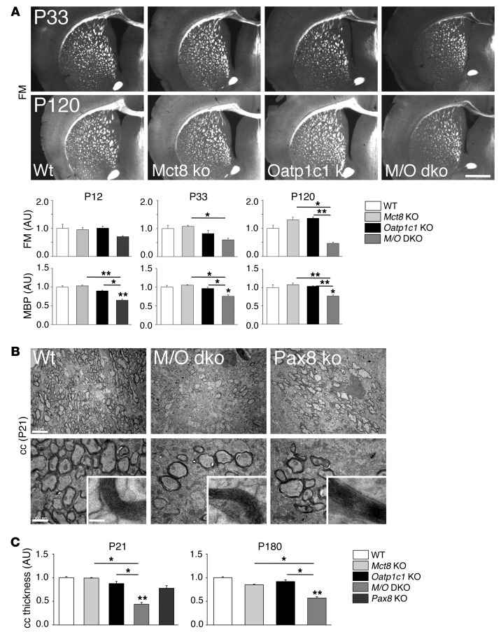
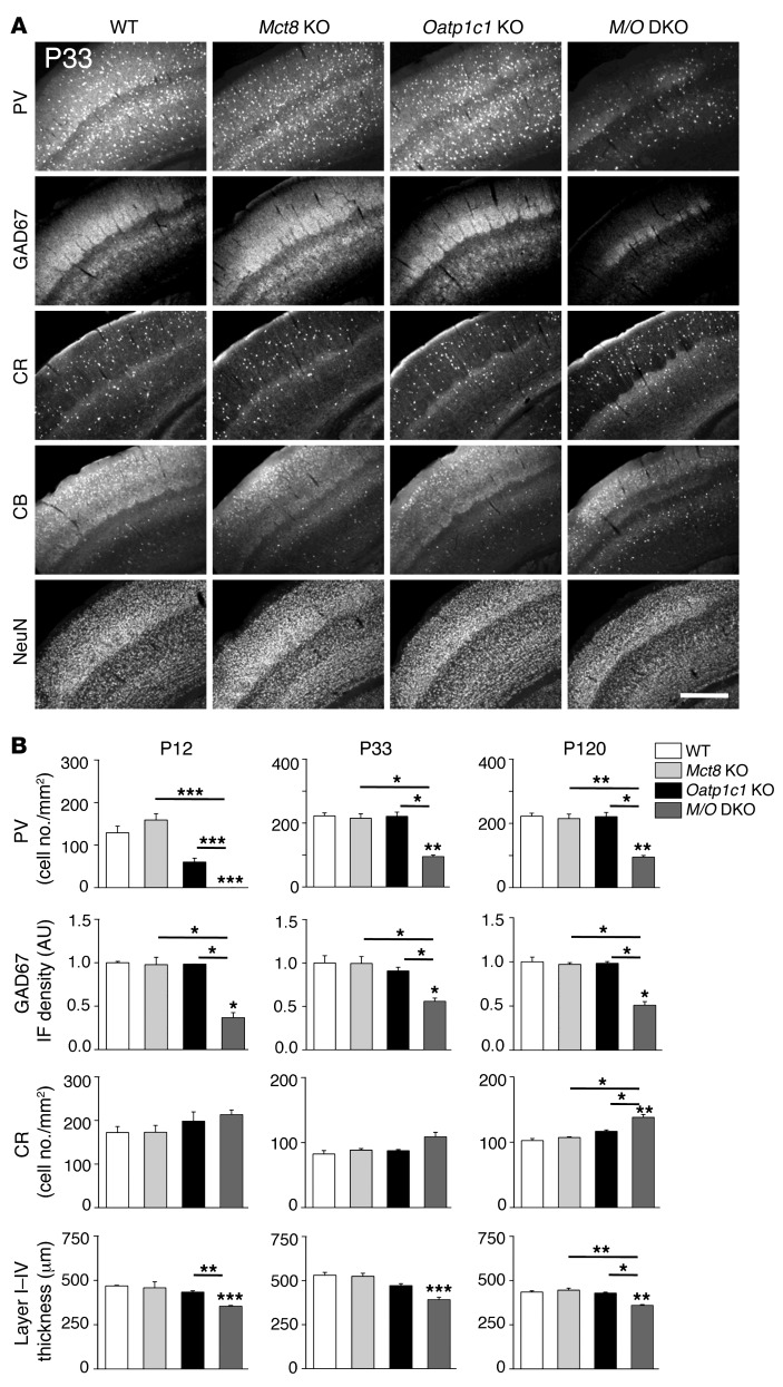
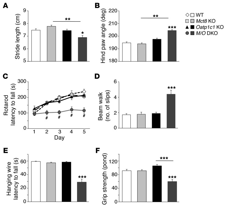
Allan-Herndon-Dudley syndrome (AHDS), a severe form of psychomotor retardation with abnormal thyroid hormone (TH) parameters, is linked to mutations in the TH-specific monocarboxylate transporter MCT8. In mice, deletion of Mct8 (Mct8 KO) faithfully replicates AHDS-associated endocrine abnormalities; however, unlike patients, these animals do not exhibit neurological impairments. While transport of the active form of TH (T3) across the blood-brain barrier is strongly diminished in Mct8 KO animals, prohormone (T4) can still enter the brain, possibly due to the presence of T4-selective organic anion transporting polypeptide (OATP1C1). Here, we characterized mice deficient for both TH transporters, MCT8 and OATP1C1 (Mct8/Oatp1c1 DKO). Mct8/Oatp1c1 DKO mice exhibited alterations in peripheral TH homeostasis that were similar to those in Mct8 KO mice; however, uptake of both T3 and T4 into the brains of Mct8/Oatp1c1 DKO mice was strongly reduced. Evidence of TH deprivation in the CNS of Mct8/Oatp1c1 DKO mice included highly decreased brain TH content as well as altered deiodinase activities and TH target gene expression. Consistent with delayed cerebellar development and reduced myelination, Mct8/Oatp1c1 DKO mice displayed pronounced locomotor abnormalities. Intriguingly, differentiation of GABAergic interneurons in the cerebral cortex was highly compromised. Our findings underscore the importance of TH transporters for proper brain development and provide a basis to study the pathogenic mechanisms underlying AHDS.


Figures

Figure 1
Figure 1. Abnormal serum TH and TSH concentrations in Mct8/Oatp1c1 DKO mice.
Serum samples of 8 animals per genotype were analyzed at P21 and revealed increased T3 and decreased T4 serum levels in Mct8/Oatp1c1 DKO mice (M/O DKO) as well as in Mct8 KO mice, whereas Oatp1c1 KO mice did not exhibit any alterations in serum TH parameters. Serum TSH concentrations were elevated in Mct8 KO mice and even more so in Mct8/Oatp1c1 DKO animals, which suggests that in the absence of both TH transporters, the HPT abnormalities characteristic of Mct8 deficiency are even more pronounced. *P < 0.05, **P < 0.01, ***P < 0.001 vs. WT, or as otherwise indicated (brackets).
Figure 2
Figure 2. Postnatal development of the hypothalamus-pituitary axis.
Brains and pituitaries from male animals (n = 4 per genotype) were collected at different postnatal time points. Hypothalamic Trh and pituitary Tshb expression were assessed by ISH. (A) Darkfield illuminations of brain sections derived from P21 animals illustrated the increase in Trh and Tshb signal intensities in Mct8 KO versus Oatp1c1 KO and WT mice. Trh expression was even further upregulated in Mct8/Oatp1c1 DKO animals. Scale bars: 250 μm (Trh), 1 mm (Tshb). (B) This optical impression was confirmed by quantification of relative signal intensities. *P < 0.05, **P < 0.01, ***P < 0.001 vs. WT, or as otherwise indicated (brackets).
Figure 3
Figure 3. Strongly diminished uptake of T4 into the brain in the absence of MCT8 and OATP1C1.
(A) For in vivo transport studies, adult animals (n = 3 per genotype and time point) were injected i.p. with 1.2 μCi [125I]T4. A blood sample was obtained, after which animals were perfused with PBS, and brains, livers, and kidneys were collected. The amount of radioactivity was expressed as percentage of the injected dose per gram of tissue. Whereas the amount of radioactivity in blood and liver samples did not differ among mutant groups, Mct8 KO and Mct8/Oatp1c1 DKO mice showed increased renal radioactivity at all analyzed time points. Most importantly, transport of radiolabeled T4 into the brain was strongly diminished in Mct8/Oatp1c1 DKO mice, whereas in the single-mutant animals, T4 uptake was reduced to approximately 50% of the respective WT levels. #P < 0.05, Mct8 KO vs. WT; §P < 0.05, Mct8/Oatp1c1 DKO vs. WT; P < 0.05, Oatp1c1 KO vs. WT. (B) Animals at P21 (n = 8 per genotype) were perfused with PBS before forebrains were collected for determining tissue TH concentrations. The concomitant inactivation of both TH transporters resulted in a pronounced decline of both T3 and T4 to approximately 10% of the levels in WT animals, which indicated that only Mct8/Oatp1c1 DKO mice exhibit a severe hypothyroid state in the CNS. ***P < 0.001 vs. WT, or as otherwise indicated (brackets).
Figure 4
Figure 4. Effect of combined MCT8 and OATP1C1 deficiency on TH metabolism in the CNS.
(A) Activities of the TH activating enzyme D2, separately measured in forebrain and cerebellar homogenates of animals at P21 (n = 6 per genotype), revealed a more than 10-fold elevation in Mct8/Oatp1c1 DKO mice, similar to that in athyroid Pax8 KO mice. (B) Determination of D3 enzymatic activities unveiled a similar reduction in Mct8 KO, Pax8 KO, and Mct8/Oatp1c1 DKO animals, whereas D3 activities were not significantly altered in Oatp1c1 KO animals. **P < 0.01, ***P < 0.001 vs. WT, or as otherwise indicated (brackets).
Figure 5
Figure 5. Consequences of combined MCT8 and OATP1C1 deficiency on T3 target gene expression in the CNS.
(A) ISH studies performed using brain sections at P21 (n = 4 per genotype) revealed for Mct8/Oatp1c1 DKO mice a strong downregulation in the signal intensities for Hr, RC3, and Aldh1a1, all known to be positively regulated by T3. Hr expression was most prominently decreased throughout the cerebral cortex, whereas RC3 expression was highly reduced in neurons of the striatum. In cortical areas, Aldh1a1-specific ISH signals were specifically reduced in astrocytes, but relatively unaltered in capillary endothelial cells. Darkfield autoradiograms also illustrated upregulation of D2 in glial cells. Interestingly, mRNA expression of Crym, an intracellular TH binding protein, was elevated specifically in the cortex and striatum. Scale bar: 600 μm (Hr, RC3, Aldh1a1, and D2); 3 mm (Crym). (B) qPCR analysis was performed using forebrain homogenates from P21 animals (n = 4–5 per genotype). Athyroid Pax8 KO mice were included and showed changes in forebrain gene expression similar to those of Mct8/Oatp1c1 DKO mice. These findings again pointed to a TH-deprived CNS in the absence of MCT8 and OATP1C1. *P < 0.05, **P < 0.01, ***P < 0.001 vs. WT.
Figure 6
Figure 6. Mct8/Oatp1c1 DKO mice display reduced myelination.
(A) Frontal vibratome brain sections of male animals (n = 3 per genotype and time point) were stained with FluoroMyelin (FM), a dye that incorporates into myelin sheaths. Additionally, consecutive sections were stained with an antibody against MBP. Quantification of the fluorescence signal densities revealed reduced staining in Mct8/Oatp1c1 DKO mice at all time points (P12, P33, and P120). Scale bar: 600 μm. (B) Ultrathin sections of the medial part of the cc were analyzed by electron microscopy at P21 (n = 3). Compared with WT animals, the number of myelinated axons was visibly decreased in Mct8/Oatp1c1 DKO mice as well as in athyroid Pax8 KO mice. However, the ultrastructure of the myelin sheaths appeared rather similar in all genotypes (higher-magnification insets). Scale bars: 5 μm (top); 500 nm (bottom); 50 nm (insets). (C) P21 and P180 coronal paraffin brain sections subjected to Gallyas silver staining were used to quantify cc thickness at the cingulum bundle (n = 3). Only Mct8/Oatp1c1 DKO mice showed decreased cc thickness at either time point. *P < 0.05, **P < 0.01, ***P < 0.001 vs. WT, or as otherwise indicated (brackets).
Figure 7
Figure 7. Histological analysis of GABAergic neurons in the somatosensory cortex.
Coronal forebrain vibratome sections from perfusion-fixed male mice (n = 3 per genotype and time point) were immunostained with antibodies recognizing the calcium-binding proteins PV, CB, and CR; the neuronal transcription factor NeuN; and the GABA-producing enzyme GAD67. (A) Representative views demonstrating visibly reduced P33, PV, and GAD67 immunoreactivity in the somatosensory cortex of Mct8/Oatp1c1 DKO mice. Scale bar: 250 μm. (B) This optical impression was confirmed by measuring GAD67 integrated fluorescence signal density and counting PV-positive cells using ImageJ. Quantification also revealed a slight increase in the number of CR-positive neurons in Mct8/Oatp1c1 DKO mice. Thickness of the cortical layers was determined in NeuN-stained brain sections and disclosed a thinner layer I–IV in Mct8/Oatp1c1 DKO mice. Overall, these data pointed to pronounced alterations in the cortical GABAergic system in Mct8/Oatp1c1 DKO animals. *P < 0.05, **P < 0.01, ***P < 0.001 vs. WT, or as otherwise indicated (brackets).
Figure 8
Figure 8. Abnormal gait and severe locomotor deficiencies in Mct8/Oatp1c1 DKO mice.
Mct8/Oatp1c1 DKO animals exhibited abnormal gait, as demonstrated by significantly reduced stride length (A), and a strongly elevated hind paw angle (B) based on footprint analysis. (C) Locomotor deficiencies were monitored by a rotarod test using 4-month-old male mice (n = 6–8 per genotype). In contrast to WT, Mct8 KO, and Oatp1c1 KO animals, Mct8/Oatp1c1 DKO mice performed poorly on the rod and did not show a learning curve. #P < 0.001 vs. WT. (D) In order to assess balance and coordination, mice were monitored while running on a beam 1 cm wide and 100 cm long. Recording the hind limb slips revealed a significantly higher number of errors only in Mct8/Oatp1c1 DKO mice. (E) Mice were further subjected to a hanging wire test in order to determine neuromuscular abnormalities. Only Mct8/Oatp1c1 DKO mice were not able to cling on a metal wire for 60 seconds. (F) As these findings point to reduced muscle performance, forepaw muscle strength was quantified using a grip strength meter, revealing an approximately 35% reduction in Mct8/Oatp1c1 DKO animals. *P < 0.05, **P < 0.01, ***P < 0.001 vs. WT, or as otherwise indicated (brackets).

References

    1. Friesema ECH, Ganguly S, Abdalla A, Manning Fox JE, Halestrap AP, Visser TJ. Identification of monocarboxylate transporter 8 as a specific thyroid hormone transporter. J Biol Chem. 2003;278(41):40128–40135. doi: 10.1074/jbc.M300909200. - DOI - PubMed
    1. Dumitrescu AM, Liao XH, Best TB, Brockmann K, Refetoff S. A novel syndrome combining thyroid and neurological abnormalities is associated with mutations in a monocarboxylate transporter gene. Am J Hum Genet. 2004;74(1):168–175. doi: 10.1086/380999. - DOI - PMC - PubMed
    1. Friesema EC, et al. Association between mutations in a thyroid hormone transporter and severe X-linked psychomotor retardation. Lancet. 2004;364(9443):1435–1437. doi: 10.1016/S0140-6736(04)17226-7. - DOI - PubMed
    1. Schwartz CE, et al. Allan-Herndon-Dudley syndrome and the monocarboxylate transporter 8 (MCT8) gene. Am J Hum Genet. 2005;77(1):41–53. doi: 10.1086/431313. - DOI - PMC - PubMed
    1. Friesema EC, Jansen J, Heuer H, Trajkovic M, Bauer K, Visser TJ. Mechanisms of disease: psychomotor retardation and high T3 levels caused by mutations in monocarboxylate transporter 8. Nat Clin Pract Endocrinol Metab. 2006;2(9):512–523. doi: 10.1038/ncpendmet0262. - DOI - PubMed

Publication types

MeSH terms

Supplementary concepts