Review
. 2014 Apr 1;6(4):a020768.
doi: 10.1101/cshperspect.a020768.

The EGFR family: not so prototypical receptor tyrosine kinases

Affiliations
Review

The EGFR family: not so prototypical receptor tyrosine kinases

Mark A Lemmon et al. Cold Spring Harb Perspect Biol. .

Abstract

The epidermal growth factor receptor (EGFR) was among the first receptor tyrosine kinases (RTKs) for which ligand binding was studied and for which the importance of ligand-induced dimerization was established. As a result, EGFR and its relatives have frequently been termed "prototypical" RTKs. Many years of mechanistic studies, however, have revealed that--far from being prototypical--the EGFR family is quite unique. As we discuss in this review, the EGFR family uses a distinctive "receptor-mediated" dimerization mechanism, with ligand binding inducing a dramatic conformational change that exposes a dimerization arm. Intracellular kinase domain regulation in this family is also unique, being driven by allosteric changes induced by asymmetric dimer formation rather than the more typical activation-loop phosphorylation. EGFR family members also distinguish themselves from other RTKs in having an intracellular juxtamembrane (JM) domain that activates (rather than autoinhibits) the receptor and a very large carboxy-terminal tail that contains autophosphorylation sites and serves an autoregulatory function. We discuss recent advances in mechanistic aspects of all of these components of EGFR family members, attempting to integrate them into a view of how RTKs in this important class are regulated at the cell surface.


Figures

Figure 1.
Figure 1.
Schematic representation of EGFR/ErbB family receptors and their ligands. (A) The domain composition of human EGFR is shown. The extracellular region contains four domains: Domain I (amino acids 1–165), domain II (amino acids 165–310), domain III (amino acids 310–480), and domain IV (amino acids 480–620). Domains I and III are closely related in sequence, as are domains II and IV. Shown are representations of the structures of domains I and IV. Domain IV contains two types of disulfide-bonded module (C1 and C2). In C1 domains, a single disulfide constrains an intervening bow-like loop. In C2 modules, two disulfides link four successive cysteines in the patterns C1–C3 and C2–C4 to give a knot-like structure. A short extracellular juxtamembrane (eJM) region separates the extracellular region from the ∼23-amino-acid transmembrane (TM) domain. Within the cell, a short intracellular juxtamembrane (iJM) region separates the tyrosine kinase domain (TKD) from the membrane. A representative EGFR TKD structure is shown. The TKD is followed by a carboxy-terminal largely unstructured tail (amino acids 953–1186) that contains at least five tyrosine autophosphorylation sites. (B) EGFR is one of four members of the EGFR/ErbB family in humans. The other members are ErbB2/HER2, for which no soluble activating ligand is shown; ErbB3/HER3, which has a significantly impaired kinase domain (Jura et al. 2009b; Shi et al. 2010); and ErbB4/HER4. The primary active moiety of the ligands for these receptors is the EGF-like domain, shown as a cartoon structure (top right). EGFR is activated by the EGFR agonists: EGF itself, TGF-α (transforming growth factor α), ARG (amphiregulin), and EGN (epigen). The bispecific ligands regulate both EGFR and ErbB4: HB-EGF (heparin-binding EGF-like growth factor), EPR (epiregulin), and BTC (betacellulin). Neuregulins (NRGs) 1 and 2 regulate ErbB3 and ErbB4, whereas NRG3 and NRG4 appear to be specific for ErbB4 (Wilson et al. 2009).
Figure 2.
Figure 2.
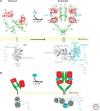
Basic model for EGF-induced dimerization and activation of EGFR. (A) In the absence of bound EGF, the human receptor is largely monomeric. The intracellular TKD is inactive, and the extracellular region adopts a “tethered” configuration in which a β-hairpin from domain II (the dimerization arm) forms intramolecular autoinhibitory interactions with domain IV. EGF binds to both domains I and III and induces a dramatic conformational change that “extends” the extracellular region and exposes the dimerization arm. With the domain II dimerization arm exposed, the EGFR extracellular region dimerizes (Burgess et al. 2003), bringing the intracellular TKDs into close proximity so that they can form the asymmetric dimer that leads to kinase activation (Zhang et al. 2006). In the asymmetric dimer, one TKD (gray) serves as the “activator,” and the other (cyan) is the “receiver” that becomes allosterically activated and trans-phosphorylates tyrosines in the tail of the activator. (B) A cartoon representation of the structural changes shown in (A). (From Ferguson 2008; adapted, with permission, from the author.)
Figure 3.
Figure 3.
Intracellular EGFR activation. (A) EGFR TKD is shown in its active (left: PDB entry 2GS6) and inactive (right: PDB entry 2GS7) configurations (Zhang et al. 2006). The amino and carboxy lobes are marked, and the nucleotide moiety is shown in stick representation. The αC helix (dark blue) occupies the “in” position in the active TKD and the “out” position in the inactive TKD. As a result, the E738 side chain is brought sufficiently close to the K721 side chain to form a salt bridge only in the active TKD. An additional short α-helix forms in the activation loop only in the inactive TKD (green) and interacts with the αC helix to promote its displacement. Mutations at two residues in this short A-loop helix (L834 and L837 in mature EGFR, L858 and L861 in pro-EGFR) are among the most common seen in EGFR-driven non-small-cell lung cancer (Sharma et al. 2007). Y845 (labeled) in the A-loop is equivalent to the key site of activating autophosphorylation in other RTKs, but its phosphorylation is not required for EGFR activation. (B) A model of an EGFR TKD asymmetric dimer, based on crystal packing in the 2GS6 structure (Zhang et al. 2006) in which an inactive TKD (from 2GS7) has been modeled in the activator position. The activator (gray) remains inactive in this discrete asymmetric dimer, and the receiver (cyan) becomes activated. (C) The same asymmetric dimer shown in (B) is shown linked to the TM domain based on a composite view of the TM and iJM structure derived from crystallographic, NMR, and computational studies (Jura et al. 2009a; Red Brewer et al. 2009; Endres et al. 2013). The TM domain forms a symmetric dimer mediated by its more amino-terminal GxxxG motif, and this, in turn, is thought to drive formation of an antiparallel dimer between helices in the amino-terminal part of the iJM region (magenta). The remainder of the iJM region in the receiver “cradles” the carboxy lobe of the activator to form the iJM “latch” that stabilizes the activating asymmetric dimer. The structure of the iJM latch region in the activator is not defined. (D) Close-up view of the iJM latch cradling the carboxy lobe of the activator TKD. Mutations at L664, V665, P675, L680, I682, and other residues in the latch impair EGFR activation, and a V665M mutation is activating (Jura et al. 2009a; Red Brewer et al. 2009).
Figure 4.
Figure 4.
Location of the small section of the EGFR carboxy-terminal tail with known structure. (A) A structure of the inactive EGFR TKD (PDB entry 1XKK) shows that two TKD-proximal regions of the carboxy-terminal tail form short helices (colored orange) that pack against the amino lobe as described in the text. Tyrosines 974 and 992 are marked, phosphorylation of which could be involved in EGFR regulation. (B) Structures of the active EGFR TKD, including this one (PDB entry 2GS6), also show the 986–994 region, including Y992, in a similar location—although not the 971–980 helix.
Figure 5.
Figure 5.
Structural basis for negative cooperativity in an EGF receptor. Negatively cooperative ligand binding is retained by the isolated extracellular region of the D. melanogaster EGFR, which binds to the EGF homolog Spitz (Spi), whereas it is lost in studies of the human receptor. Crystallographic studies have revealed the structural basis for this negative cooperativity. The D. melanogaster EGFR extracellular region does not form the tethered structure depicted in Figure 2A, remaining as an untethered monomer or forming a symmetric (crystallographic) dimer even in the absence of bound ligand (Alvarado et al. 2009). After binding of ligand to the left-hand molecule in the symmetric dimer, domains I and III are wedged apart as described in the text so that domain II of the left-hand molecule “collapses” onto its counterpart in the right-hand molecule. The result is a singly ligated asymmetric dimer. This transition restrains the second ligand-binding site so that the wedging apart of domains I and III is disfavored, reducing the affinity of ligand for the second site—resulting in negative cooperativity. The structures in the upper panel are actual crystal structures (PDB entries 3I2T, 3LTG, and 3LTF) and are redrawn as cartoons in the lower part of the figure for clarity (Alvarado et al. 2009, 2010).

References

    1. Adrain C, Freeman M 2014. Regulation of receptor tyrosine kinase ligand processing. Cold Spring Harb Perspect Biol 10.1101/cshperspect.a008995 - DOI - PMC - PubMed
    1. Alvarado D, Klein DE, Lemmon MA 2009. ErbB2 resembles an autoinhibited invertebrate epidermal growth factor receptor. Nature 461: 287–291 - PMC - PubMed
    1. Alvarado D, Klein DE, Lemmon MA 2010. Structural basis for negative cooperativity in growth factor binding to an EGF receptor. Cell 142: 568–579 - PMC - PubMed
    1. Alvarez CV, Shon KJ, Miloso M, Beguinot L 1995. Structural requirements of the epidermal growth factor receptor for tyrosine phosphorylation of eps8 and eps15, substrates lacking Src SH2 homology domains. J Biol Chem 270: 16271–16276 - PubMed
    1. Arndt-Jovin DJ, Botelho MG, Jovin TM 2014. Structure–function relationships of ErbB RTKs in the plasma membranes of living cells. Cold Spring Harb Perspect Biol 10.1101/cshperspect.a008961 - DOI - PMC - PubMed