. 2014 May;42(9):5702-14.
doi: 10.1093/nar/gku228. Epub 2014 Apr 1.

Cellular location and activity of Escherichia coli RecG proteins shed light on the function of its structurally unresolved C-terminus

Affiliations

Cellular location and activity of Escherichia coli RecG proteins shed light on the function of its structurally unresolved C-terminus

Amy L Upton et al. Nucleic Acids Res. 2014 May.

Abstract

RecG is a DNA translocase encoded by most species of bacteria. The Escherichia coli protein targets branched DNA substrates and drives the unwinding and rewinding of DNA strands. Its ability to remodel replication forks and to genetically interact with PriA protein have led to the idea that it plays an important role in securing faithful genome duplication. Here we report that RecG co-localises with sites of DNA replication and identify conserved arginine and tryptophan residues near its C-terminus that are needed for this localisation. We establish that the extreme C-terminus, which is not resolved in the crystal structure, is vital for DNA unwinding but not for DNA binding. Substituting an alanine for a highly conserved tyrosine near the very end results in a substantial reduction in the ability to unwind replication fork and Holliday junction structures but has no effect on substrate affinity. Deleting or substituting the terminal alanine causes an even greater reduction in unwinding activity, which is somewhat surprising as this residue is not uniformly present in closely related RecG proteins. More significantly, the extreme C-terminal mutations have little effect on localisation. Mutations that do prevent localisation result in only a slight reduction in the capacity for DNA repair.


Figures

Figure 1.
Figure 1.
A chromosomal copy of eYFP-recG confers resistance to mitomycin C and UV light. The strains used are identified in parentheses.
Figure 2.
Figure 2.
Cellular localisation of RecG. (A) RecG co-localises with SeqA. The strains identified in parentheses carry a construct expressing both eYFP-RecG and eCFP-SeqA. The panels show phase contrast images merged with the indicated fluorescence images. (B) Co-localisation depends upon the C-terminus of RecG. The panels show merged phase contrast and fluorescence images, with the constructs identified underneath. (C) Identification and mutation of conserved residues within C-terminus of RecG. (i) Multiple alignment of C-terminal sequences of RecG proteins. The sequences are from RecG proteins that have a C-terminal region of similar length. Shading is related directly to amino acid similarity. Residues are numbered according to E. coli RecG. The structure associated with residues in the region corresponding to the last 20 amino acids (marked by a black line above the E. coli sequence) has not been resolved at the atomic level (22). (ii) Schematic representation of the major C-terminal RecG deletions and substitutions used.
Figure 3.
Figure 3.
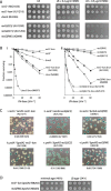
Effect of RecG C5 and C15 deletions and RW substitutions on sensitivity to DNA damage and viability. (A) Sensitivity to mitomycin C. (B) Sensitivity to UV light. The strains used were as in (A) plus additional constructs identified in parentheses. Data for strain AU1232 are duplicated in panels (i) and (ii) for the purposes of comparison. (C) Synthetic lethality assays illustrating the effect of the C-terminal RecG mutations on the viability of ΔrnhA and ΔpolA2 strains on LB agar. The relevant genotype is shown above each photograph, with the strain number shown in parentheses. The fraction of white colonies is shown below, with the number of white colonies/total colonies analysed shown in parentheses. (D) Relative plating efficiencies of ΔpolA2 recG+-kan and ΔpolA2 recG[RW] cells. Cultures of the strains identified were grown in 56/2 glucose minimal salts medium to an A650 of 0.48, diluted in 10-fold steps from 10−1 to 10−5, and 10 μl aliquots spotted on minimal and LB agar, as indicated. Plates were photographed after incubation for 24 h (LB agar) or 48 h (minimal agar).
Figure 4.
Figure 4.
Effect of extreme RecG C-terminal mutations on sensitivity to DNA damage. (A) Sensitivity to mitomycin C. The strains used are identified in parentheses. (B) Sensitivity to UV light. The strains used were as in (A) plus additional constructs identified in parentheses. Data for strains AU1218, AU1232, N4256 and N4971 are reproduced from Figure 3B for the purposes of comparison.
Figure 5.
Figure 5.
Effect of extreme RecG C-terminal mutations on the viability of ΔrnhA and ΔpolA2 cells. (A) and (B) Synthetic lethality assays. The relevant genotype is shown above each photograph, with the strain number shown in parentheses. The fraction of white colonies is shown below, with the number of white colonies/total colonies analysed shown in parentheses. (A) Assays with ΔrnhA constructs on LB indicator plates. The data for strain AU1217 are reproduced from Figure 3C(i) for comparison. (B) Assays with ΔpolA2 constructs on both LB (top row) and 56/2 glucose minimal salts indicator plates. Panel (iv) illustrates the formation of large colonies by faster growing variants (suppressors) accumulating in the white colonies shown in panel (iii). (C) Low viability of recG[ΔC1] ΔpolA2 cells on LB agar. Three independent colonies of plasmid-free recG[ΔC1] ΔpolA2 cells established on 56/2 glucose minimal salts indicator plates were inoculated into 56/2 glucose minimal salts and incubated until the cell density reached an A650 of 0.48. Samples were then diluted in 56/2 salts in 10-fold steps from 10−1 to 10−5, before spotting 10 μl aliquots of each dilution on 56/2 glucose minimal salts agar and LB agar, as indicated. These plates were photographed after 24 and 48 h incubation, respectively.
Figure 6.
Figure 6.
DNA binding and unwinding activities of RecG C-terminal mutants. (A) DNA binding assays. The autoradiograph shown is of representative band-shift assays with Holliday junction substrate J12. Each set of reactions from left to right used the RecG proteins indicated at 0, 0.1, 0.4, 1.6, 6.4, 25 and 100 nM and 32P-labelled J12 DNA at 0.2 nM. (B) DNA unwinding assays. The graphs show the rates of dissociation of 32P-labelled Holliday junction and replication fork substrates by the proteins indicated. Reactions contained substrate DNA at 0.2 nM and RecG protein at 0.5 nM. Data are means (±SE) of at least three independent experiments.

References

    1. Lloyd R.G., Buckman C. Genetic analysis of the recG locus of Escherichia coli K-12 and of its role in recombination and DNA repair. J. Bacteriol. 1991;173:1004–1011. - PMC - PubMed
    1. Rudolph C.J., Upton A.L., Briggs G.S., Lloyd R.G. Is RecG a general guardian of the bacterial genome? DNA Repair (Amst.) 2010;9:210–223. - PubMed
    1. Rocha E.P., Cornet E., Michel B. Comparative and evolutionary analysis of the bacterial homologous recombination systems. PLoS Genet. 2005;1:e15. - PMC - PubMed
    1. Sharples G.J., Ingleston S.M., Lloyd R.G. Holliday junction processing in bacteria: insights from the evolutionary conservation of RuvABC, RecG, and RusA. J. Bacteriol. 1999;181:5543–5550. - PMC - PubMed
    1. Lloyd R.G. Conjugational recombination in resolvase-deficient ruvC mutants of Escherichia coli K-12 depends on recG. J. Bacteriol. 1991;173:5414–5418. - PMC - PubMed

Publication types