. 2014 May;42(9):5846-62.
doi: 10.1093/nar/gku237. Epub 2014 Apr 1.

Human MUS81-EME2 can cleave a variety of DNA structures including intact Holliday junction and nicked duplex

Affiliations

Human MUS81-EME2 can cleave a variety of DNA structures including intact Holliday junction and nicked duplex

Tamir Amangyeld et al. Nucleic Acids Res. 2014 May.

Abstract

MUS81 shares a high-degree homology with the catalytic XPF subunit of the XPF-ERCC1 endonuclease complex. It is catalytically active only when complexed with the regulatory subunits Mms4 or Eme1 in budding and fission yeasts, respectively, and EME1 or EME2 in humans. Although Mus81 complexes are implicated in the resolution of recombination intermediates in vivo, recombinant yeast Mus81-Mms4 and human MUS81-EME1 isolated from Escherichia coli fail to cleave intact Holliday junctions (HJs) in vitro. In this study, we show that human recombinant MUS81-EME2 isolated from E. coli cleaves HJs relatively efficiently, compared to MUS81-EME1. Furthermore, MUS81-EME2 catalyzed cleavage of nicked and gapped duplex deoxyribonucleic acids (DNAs), generating double-strand breaks. The presence of a 5' phosphate terminus at nicks and gaps rendered DNA significantly less susceptible to the cleavage by MUS81-EME2 than its absence, raising the possibility that this activity could play a role in channeling damaged DNA duplexes that are not readily repaired into the recombinational repair pathways. Significant differences in substrate specificity observed with unmodified forms of MUS81-EME1 and MUS81-EME2 suggest that they play related but non-overlapping roles in DNA transactions.


Figures

Figure 1.
Figure 1.
Purification of the recombinant human MUS81-EME2 complex from E. coli extracts that contains intrinsic endonuclease activity which can cleave intact Holliday junction (iHJ). (A) The purification scheme used to purify the recombinant human MUS81-EME2 complex. The recombinant human MUS81-EME2 was expressed in E. coli and purified using the same procedure as reported (20,32), but modified by the introduction of two additional purification steps (see text for details). P11, Ni2+-NTA and heparin indicate phosphocellulose, Ni2+-NTA affinity and heparin column chromatography, respectively. IDZ, imidazole. Glycerol gradient indicates the final purification procedure using glycerol gradient sedimentation. Fraction I is the crude extract prepared from E. coli carrying the bicistronic vector pET21d-MUS81/His-EME2 (20), and other fractions from each step are as indicated. (B) An aliquot (250 μl) of Fraction IV from heparin column chromatography was subjected to glycerol gradient sedimentation and the resulting fractions were analyzed in a 10% SDS-PAGE and the gel was stained with Coomassie Brilliant Blue R-250. The sizes of molecular mass markers are indicated in kDa. The positions of MUS81 and EME2 are as indicated. LO, load-on. (C) and (D) A fixed amount (15 fmol) of intact iHJ [panel (C)] and 3′ flap (3′F) [panel (D)] substrates was incubated with 0.02 μl (1 μl of 50-fold dilution) of the indicated glycerol gradient fractions of MUS81-EME2 at 37°C for 30 min. The schematic structures of the substrate and marker DNA are shown at the right side of the gel. The DNA substrates used were prepared as described in the Materials and Methods section. Asterisks indicate the position of the 5′-32P label. Two marker DNAs (2-nt gapped 50-bp linear and 25-bp linear duplex DNA) were used as indicated to monitor the migration of DNA products. Reaction products were subjected to 10% polyacrylamide gel electrophoresis (PAGE) in 1X TBE at 130 V. The amount of cleavage product formed is indicated at the bottom of the gel. LO, load-on (Fraction IV in Figure 1A).
Figure 2.
Figure 2.
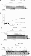
Comparison of endonuclease activities of human MUS81-EME1 and MUS81-EME2 complexes. Standard endonuclease assays were performed with 15 fmol of 3′F (A) and (B), nDS (C) and iHJ (D) substrates in the presence of varying amounts (10, 20 or 40 fmol) of MUS81-EME1 and MUS81-EME2 complexes. Assays were performed at 37°C and reactions were stopped at the indicated time points (1, 2, 4, 8, 15 and 30 min). Addition or omission of MUS81-EME2 is indicated by ‘+’ or ‘−’, respectively. DNA substrates and the marker DNAs are shown at the right side of the gel. The amount of cleavage product formed is indicated at the bottom of the gel. Asterisks indicate the position of the 5′-32P label. (B)–(D) The amount of cleavage products was plotted against the time of incubation.
Figure 3.
Figure 3.
Human MUS81-EME2 processed different RF structures. (A) Standard endonuclease assays were performed with 15 fmol of 3′F, RF, Rle and Rla substrates in the presence of increasing amount (5, 10 and 20 fmol) of MUS81 complexes. The substrates used are indicated at the top of the gel. Asterisks indicate the position of the 5′-32P label. Reaction mixtures were incubated at 37°C for 30 min. (B) The same reaction as in panel (A) was repeated with differently labeled Rla and Rle substrates. Rla and Rle substrates (10 fmol) were incubated with increasing amounts (1, 5 and 25 fmol) of MUS81-EME2 at 37°C for 60 min. Omission of protein is indicated by ‘−’. The structures of the DNA substrates used are shown at the top of the gel. Shown at the left and right of the gel are the structures of cleavage products. The amount of cleavage products is indicated at the bottom of the gel. The primary and secondary cleavage sites are indicated by the solid and open arrows, respectively, for each substrate used. (C) The reaction mixtures incubated with 25 fmol of MUS81-EME2 and each substrate as indicated above the gel were stopped by the addition of 2X stop solution (95% formamide, 20-mM EDTA, 0.1% bromophenol blue and 0.1% xylene cyanol) after 60 min of incubation at 37°C, and cleavage products were subjected to 15% denaturing PAGE containing 7-M urea. The molecular size markers are synthetic oligonucleotides labeled at their 5′ ends, and their size (nt) is indicated.
Figure 4.
Figure 4.
Analysis of cleavage of each strand in an nDS DNA substrate by human MUS81-EME2. (A) Reaction mixtures containing 15 fmol of one of the nDS (nDS1, nDS2 and nDS3) substrates and increasing amounts (5, 10, 20 and 40 fmol) of MUS81-EME2 were incubated at 37°C for 30 min. The cleavage products were subjected to 15% denaturing PAGE. The molecular mass markers are as described in Figure 4C. Asterisks indicate the position of the 32P-label.(B) The cleavage sites of MUS81-EME2 on the nDS substrate are indicated by arrows; longer arrows indicate more preferred cleavage sites.
Figure 5.
Figure 5.
The presence or absence of 5′ phosphate at a nick influences the cleavage of duplex DNA by MUS81-EME2. (A) Endonuclease assays were performed using 15 fmol of unligatable (no phopshpate residue at the nick) or ligatable (+5′ phosphate at the nick) nDS and 30 fmol of MUS81-EME2. The structures of DNA substrates are shown at the top of the gel. An open circle indicates a phosphoryl group. Reactions were stopped at the indicated time points (1, 2, 4, 8, 15 and 30 min). (B) The amount of cleavage products formed in panel (A) was plotted against incubation time. (C) The same reaction as in panel (A) was repeated with 15 fmol of 3′F with and without a phosphoryl group at the 5′ end at the nick of junction. The structures of DNA substrates used are shown at the top of the gel. An open circle indicates a phosphoryl group. (D) Endonuclease assays were performed using a fixed amount (30 fmol) of MUS81-EME2 and 15 fmol of gapped duplexes (gDS) of varying length (1–10 nt) of gap, with or without a 5′ phosphate at the 5′ end of the downstream oligonucleotide. The structure of the gDS substrate is shown at the top of the gel. X indicates the upstream strand of varying size. The gap size of each substrate used is as indicated. Addition or omission of MUS81-EME2 and the 5′ phosphoryl group (5′ PO4) are indicated by ‘+’ or ‘−’, respectively. The marker DNA (25-bp linear duplex) is shown at the right side of the gel. The amount of cleavage products formed is indicated at the bottom of the gel. Asterisks indicate the position of the 5′-32P label.
Figure 6.
Figure 6.
Analysis of HJ cleavage activity of recombinant MUS81-EME2. (A) The structures of DNA substrates used are shown at the top of the gel. An open circle indicates a phosphoryl group. Three substrates including iHJ, nHJ (no 5′ PO4) and nHJ (+5′ PO4) (15 fmol each) were incubated with increasing amounts (2.5, 5, 10 and 20 fmol) of MUS81-EME2 and the products analyzed as above. Marker DNAs are shown at the right side of the gel. Asterisks indicate the position of the 5′-32P label. The amount of cleavage products is indicated at the bottom of the gel. (B) The amount of cleavage products formed in panel (A) was plotted against the amount of MUS81-EME2 added. (C) The same reactions as in panel (B) were repeated and products were subjected to 15% denaturing PAGE. The molecular mass markers are as described above. Omission of MUS81-EME2 is indicated by ‘−’.
Figure 7.
Figure 7.
Cleavage sites of human MUS81-EME2 on iHJ. (A) Endonuclease assays were performed as described in the Materials and Methods section using four differently labeled iHJ substrates (iHJ1, iHJ2, iHJ3 and iHJ4). Reaction mixtures containing 10 fmol of each iHJ substrate and increasing amounts (10, 30 and 90 fmol) of MUS81-EME2 were incubated for 60 min at 37°C. DNA substrates used are shown at the top of the gel. The three marker DNAs (2-nt gapped 50-bp linear duplex, 25-bp linear duplex and 25-nt ssDNA) are shown at the right side of the gel. Asterisks indicate the position of the 5′-32P label. The amount of cleavage products is indicated at the bottom of the gel. (B) The amount of cleavage products formed in panel (A) was plotted against the amount of MUS81-EME2 added. (C) The products were subjected to 15% denaturing PAGE. The molecular mass markers are synthetic oligonucleotides labeled at their 5′ ends, and size (nt) is indicated. (D) The sites on the iHJ substrate cleaved by MUS81-EME2 are indicated by arrows. Longer arrows indicate preferred cleavage sites.
Figure 8.
Figure 8.
The C-terminal EME1 fragment does not substitute for EME2. Endonuclease assays were performed as described in the Materials and Methods section using nHJ (A) and iHJ (C) substrates. A fixed amount (15 fmol) of DNA substrate was incubated for 60 min at 37°C with increasing amounts (12, 25 and 50 fmol) of MUS81-EME1(244–583), MUS81-EME1 and MUS81-EME2. DNA substrates used are shown at the top of the gel. The amount of cleavage products is indicated at the bottom of the gel. (B) and (D) The amount of cleavage products formed in panels (A) and (C), respectively, was plotted against the amount of each MUS81 complex used.

References

    1. Kuzminov A. Collapse and repair of replication forks in. Escherichia Coli Mol. Microbiol. 1995;16:373–384. - PubMed
    1. Fabre F., Chan A., Heyer W.D., Gangloff S. Alternate pathways involving Sgs1/Top3, Mus81/Mms4, and Srs2 prevent formation of toxic recombination intermediates from single-stranded gaps created by DNA replication. Proc. Natl. Acad. Sci. U.S.A. 2002;99:16887–16892. - PMC - PubMed
    1. Cox M.M., Goodman M.F., Kreuzer K.N., Sherratt D.J., Sandlerk S.J., Marians K.J. The importance of repairing stalled replication forks. Nature. 2000;404:37–41. - PubMed
    1. Kowalczykowski S.C. Initiation of genetic recombination and recombination-dependent replication. Trends Biochem. Sci. 2000;25:156–165. - PubMed
    1. Higgins N.P., Kato K., Strauss B. A model for replication repair in mammalian cells. J. Mol. Biol. 1976;101:417–425. - PubMed

Publication types