Mycotic abdominal aortic aneurysm caused by bacteroides thetaiotaomicron and acinetobacter lwoffii: the first case in Korea
- PMID: 24693472
- PMCID: PMC3970306
- DOI: 10.3947/ic.2014.46.1.54
Mycotic abdominal aortic aneurysm caused by bacteroides thetaiotaomicron and acinetobacter lwoffii: the first case in Korea
Abstract
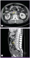
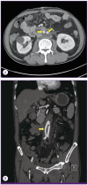
Mycotic aneurysms are uncommon, but are fatal without appropriate management. Previous reports have shown that anaerobes and gram-negative organisms are less common but more dangerous than other causative agents of mycotic aneurysm. We report the case of a 60-year-old man with poorly controlled diabetes mellitus and atherosclerosis in the aorta, and a 10-day of history of lower abdominal pain and fever. This man was diagnosed with an uncommon abdominal aorta mycotic aneurysm caused by Bacteroides thetaiotaomicron and Acinetobacter lwoffii. The aneurysm was successfully treated with antibiotics therapy and aorto-bi-external iliac artery bypass with debridement of the infected aortic wall. We present this case together with a review of the relevant literature.
Keywords: Acinetobacter lwoffii; Aneurysm; Bacteroides thetaiotaomicron; Infected.
Figures


References
-
- Malouf JF, Chandrasekaran K, Orszulak TA. Mycotic aneurysms of the thoracic aorta: a diagnostic challenge. Am J Med. 2003;115:489–496. - PubMed
-
- Müller BT, Wegener OR, Grabitz K, Pillny M, Thomas L, Sandmann W. Mycotic aneurysms of the thoracic and abdominal aorta and iliac arteries: experience with anatomic and extra-anatomic repair in 33 cases. J Vasc Surg. 2001;33:106–113. - PubMed
-
- Johnson JR, Ledgerwood AM, Lucas CE. Mycotic aneurysm. New concepts in therapy. Arch Surg. 1983;118:577. - PubMed
LinkOut - more resources
Full Text Sources
Other Literature Sources

