. 2014 Apr 2:14:87.
doi: 10.1186/1471-2229-14-87.

The onset of grapevine berry ripening is characterized by ROS accumulation and lipoxygenase-mediated membrane peroxidation in the skin

Affiliations

The onset of grapevine berry ripening is characterized by ROS accumulation and lipoxygenase-mediated membrane peroxidation in the skin

Stefania Pilati et al. BMC Plant Biol. .

Abstract

Background: The ripening of fleshy fruits is a complex developmental program characterized by extensive transcriptomic and metabolic remodeling in the pericarp tissues (pulp and skin) making unripe green fruits soft, tasteful and colored. The onset of ripening is regulated by a plethora of endogenous signals tuned to external stimuli. In grapevine and tomato, which are classified as non-climacteric and climacteric species respectively, the accumulation of hydrogen peroxide (H2O2) and extensive modulation of reactive oxygen species (ROS) scavenging enzymes at the onset of ripening has been reported, suggesting that ROS could participate to the regulatory network of fruit development. In order to investigate this hypothesis, a comprehensive biochemical study of the oxidative events occurring at the beginning of ripening in Vitis vinifera cv. Pinot Noir has been undertaken.

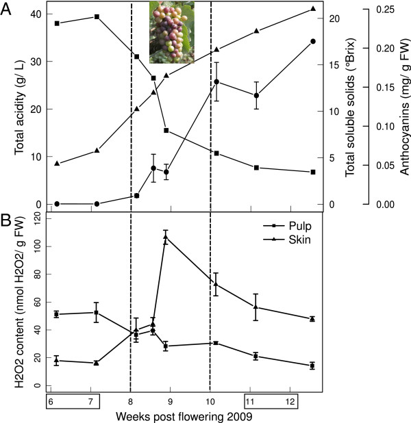
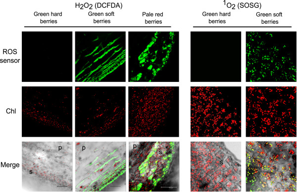
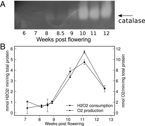
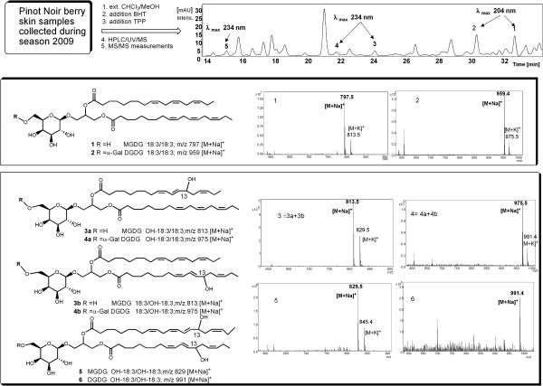
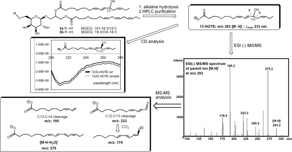
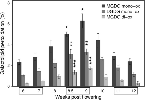
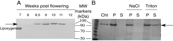
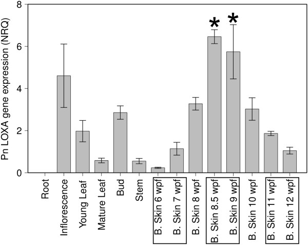
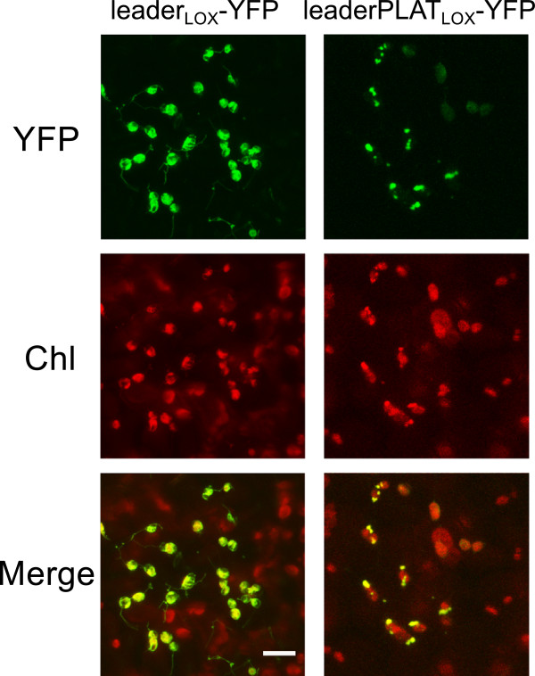
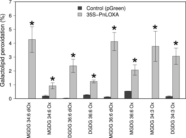
Results: ROS-specific staining allowed to visualize not only H2O2 but also singlet oxygen (1O2) in berry skin cells just before color change in distinct subcellular locations, i.e. cytosol and plastids. H2O2 peak in sample skins at véraison was confirmed by in vitro quantification and was supported by the concomitant increase of catalase activity. Membrane peroxidation was also observed by HPLC-MS on galactolipid species at véraison. Mono- and digalactosyl diacylglycerols were found peroxidized on one or both α-linolenic fatty acid chains, with a 13(S) absolute configuration implying the action of a specific enzyme. A lipoxygenase (PnLOXA), expressed at véraison and localizing inside the chloroplasts, was indeed able to catalyze membrane galactolipid peroxidation when overexpressed in tobacco leaves.

Conclusions: The present work demonstrates the controlled, harmless accumulation of specific ROS in distinct cellular compartments, i.e. cytosol and chloroplasts, at a definite developmental stage, the onset of grape berry ripening. These features strongly candidate ROS as cellular signals in fruit ripening and encourage further studies to identify downstream elements of this cascade. This paper also reports the transient galactolipid peroxidation carried out by a véraison-specific chloroplastic lipoxygenase. The function of peroxidized membranes, likely distinct from that of free fatty acids due to their structural role and tight interaction with photosynthesis protein complexes, has to be ascertained.


Figures

Figure 1
Figure 1
H2O2 content and biochemical changes in Pinot Noir berries during development. A: Mean values of total acids (squares, expressed as grams of tartaric acid per liter) and sugars (triangles, expressed as total soluble solids in °Brix) of the must obtained from three clusters, at each time point. Berry skins anthocyanin content (circles) is expressed as grams of pelargonidin-3-glucoside per gram of berry fresh weight. B: H2O2 was measured separately in skin and pulp tissues of sampled berries. Data are means of three biological replicates ± se. The x-axis represents time in weeks post flowering (wpf). Véraison is indicated between dashed lines (8-10 wpf). Pre- (6-7 wpf) and post-véraison (11-12 wpf) stages are indicated by boxes. The picture of a cluster at mid-véraison shows the typical asynchrony of berries at this developmental transition.
Figure 2
Figure 2
Confocal images of Pinot Noir berries (100-μm sections) sampled at the green hard, green soft and pale red stages, stained for H2O2and1O2. The sections were incubated with either 30 μM DCFDA or 30 μM SOSG (ROS sensors). Chlorophyll fluorescence has been recorded (Chl) to localize chloroplasts inside the cells. Merge is the computed overlay of the two fluorescence images and the bright field. Reference bars are 75 μm for H2O2 imaging and 25 μm for 1O2. Skin and pulp are indicated in the merge pictures with a “s” and “p”, respectively. For 1O2 imaging, only skin is visualized, at a higher magnification.
Figure 3
Figure 3
Catalase activity during Pinot Noir berry development. Native protein lysates were obtained from berry skins sampled at the indicated time points. A: Zymogram of catalase activity using 50 μg total proteins per lane. B: Catalase specific activity measured in vitro by determining either H2O2 consumption (absorbance at 240 nm) or O2 production (in-line O2 recording using direct injection MS). Data are means of biological duplicates ± se.
Figure 4
Figure 4
Overview of the characterization study of galactolipids extracted from Pinot Noir berry skins at véraison (9 wpf). The chromatogram shows eluted peaks recorded at 210 nm, with retention time shown on the x-axis. The mass spectra of the indicated peaks revealed that peaks 1 and 2 are attributable to MGDG 36:6 and DGDG 36:6. Peaks 3 and 4 are attributable to the corresponding mono-oxidized forms and peak 5 to the di-oxidized MGDG 36:6. Di-oxidized DGDG 36:6 has been identified but was barely detectable in the chromatogram.
Figure 5
Figure 5
Overview of the characterization study of the oxidized fatty acid chains obtained after hydrolysis of oxidized MGDG 36:6. ESI MS/MS has been performed to assess the regiospecificity of the oxidation event and CD analysis has been performed to define its stereospecificity.
Figure 6
Figure 6
Galactolipid peroxidation profiles during Pinot Noir berry development. The mono-oxidized and di-oxidized forms of MGDG 36:6 and DGDG 36:6 are shown as percentage of total MGDG and DGDG, respectively. Data are means of three biological replicates ± sd. Lipid peroxidation at pre-véraison (6 and 7 wpf), véraison (8.5 and 9 wpf) and ripening (11 and 12 wpf) were analyzed by ANOVA and Tukey’s HSD (honestly significant difference) test. Asterisks indicate that the amount of peroxidized species accumulated at véraison is significantly different from that of the other two moments (p < 0.01).
Figure 7
Figure 7
Western blot analysis of lipoxygenase expression in Pinot Noir berry skin extracts. A: Analysis of plastid lipoxygenases expression during berry development using a commercial antibody against Arabidopsis LOX2 and 10 μg of total protein extracts per lane. B: Analysis of plastid lipoxygenase expression in chloroplast-enriched samples obtained from fresh berry skins collected at 9 wpf. Total chloroplast protein extract (Chl) was fractionated into membrane (P) and soluble (S) fractions by centrifugation. Membrane pellets were treated with 1 M NaCl or 0.05% Triton X-100, incubated for 10 min on ice and centrifuged again to separate the membrane (P) and the soluble (S) fractions. Pellets were resuspended in a volume identical to the corresponding soluble fractions and loaded in equal amounts for separation by SDS-PAGE and detection by western blot. MW markers: molecular weight markers (kDa).
Figure 8
Figure 8
PnLOXA gene expression in grapevine tissues and in berry skins along development (6–12 wpf). Normalized relative quantities ± se were calculated using three reference genes; n = 3. PnLOXA expression at véraison (marked by asterisks) was significantly different from pre-véraison (6-7 wpf) and ripening (11-12 wpf) as assessed by ANOVA and Tukey HSD test (p < 0.01).
Figure 9
Figure 9
PnLOXA localization demonstrated by the expression of YFP fusion constructs in grapevine leaves. Leaves were infiltrated with Agrobacterium tumefaciens carrying the pGreen[PnLOXAtransitpeptide1-47-YFP] and pGreen[PnLOXAtransitpeptidePLAT1-220-YFP] constructs. Chlorophyll (Chl) and YFP fluorescence were recorded using Leica SP II confocal microscope. Merge is the computed overlay of the two fluorescence images. Reference bar is 10 μm.
Figure 10
Figure 10
Galactolipid analysis of tobacco leaves transiently expressing PnLOXA. Leaves transformed either with the PnLOXA or the empty vector (pGreen) as control were collected 7 days after Agrobacterium inoculation. Galactolipid peroxidation is reported as a percentage of mono- and di-oxidized species within each class, normalized on the amount of PnLOXA protein. Data are means of three replicates ± sd. ANOVA and Tukey HSD test were performed to compare control and PnLOXA over-expressing samples. Asterisks indicate significant differences from control at p < -0.05.

References

    1. Terrier N, Glissant D, Grimplet J, Barrieu F, Abbal P, Couture C, Ageorges A, Atanassova R, Léon C, Renaudin J-P, Dédaldéchamp F, Romieu C, Delrot S, Hamdi S. Isogene specific oligo arrays reveal multifaceted changes in gene expression during grape berry (Vitis vinifera L.) development. Planta. 2005;222:832–847. doi: 10.1007/s00425-005-0017-y. - DOI - PubMed
    1. Deluc LG, Grimplet J, Wheatley MD, Tillett RL, Quilici DR, Osborne C, Schooley DA, Schlauch KA, Cushman JC, Cramer GR. Transcriptomic and metabolite analyses of Cabernet Sauvignon grape berry development. BMC Genomics. 2007;8:429. doi: 10.1186/1471-2164-8-429. - DOI - PMC - PubMed
    1. Pilati S, Perazzolli M, Malossini A, Cestaro A, Demattè L, Fontana P, Dal Ri A, Viola R, Velasco R, Moser C. Genome-wide transcriptional analysis of grapevine berry ripening reveals a set of genes similarly modulated during three seasons and the occurrence of an oxidative burst at vèraison. BMC Genomics. 2007;8:428. doi: 10.1186/1471-2164-8-428. - DOI - PMC - PubMed
    1. Lund S, Peng F, Nayar T, Reid K, Schlosser J. Gene expression analyses in individual grape (Vitis vinifera L.) berries during ripening initiation reveal that pigmentation intensity is a valid indicator of developmental staging within the cluster. Plant Mol Biol. 2008;68:301–315. doi: 10.1007/s11103-008-9371-z. - DOI - PubMed
    1. Zenoni S, Ferrarini A, Giacomelli E, Xumerle L, Fasoli M, Malerba G, Bellin D, Pezzotti M, Delledonne M. Characterization of transcriptional complexity during berry development in Vitis vinifera using RNA-Seq1[W] Plant Physiol. 2010;152:1787–1795. doi: 10.1104/pp.109.149716. - DOI - PMC - PubMed

Publication types

MeSH terms

LinkOut - more resources