. 2014 Apr 3:14:90.
doi: 10.1186/1471-2229-14-90.

Galactosyltransferases from Arabidopsis thaliana in the biosynthesis of type II arabinogalactan: molecular interaction enhances enzyme activity

Affiliations

Galactosyltransferases from Arabidopsis thaliana in the biosynthesis of type II arabinogalactan: molecular interaction enhances enzyme activity

Adiphol Dilokpimol et al. BMC Plant Biol. .

Abstract

Background: Arabinogalactan proteins are abundant proteoglycans present on cell surfaces of plants and involved in many cellular processes, including somatic embryogenesis, cell-cell communication and cell elongation. Arabinogalactan proteins consist mainly of glycan, which is synthesized by post-translational modification of proteins in the secretory pathway. Importance of the variations in the glycan moiety of arabinogalactan proteins for their functions has been implicated, but its biosynthetic process is poorly understood.

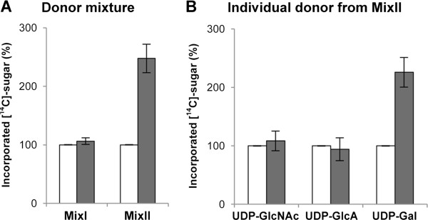
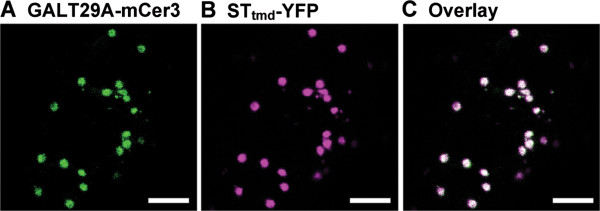
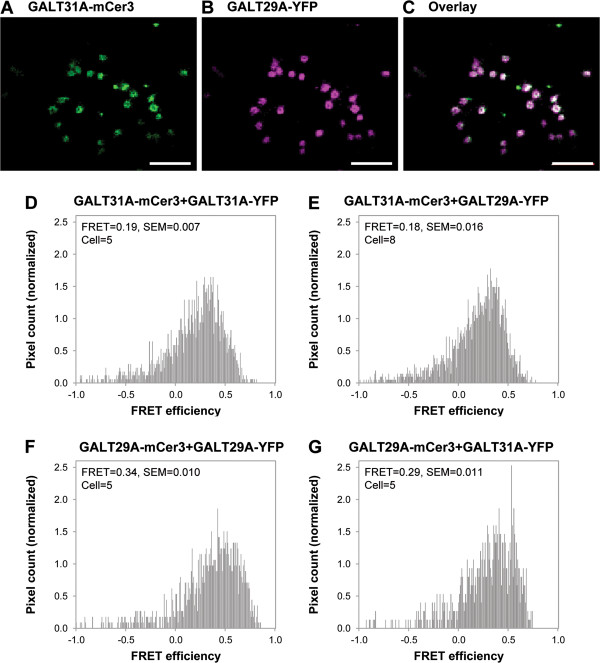
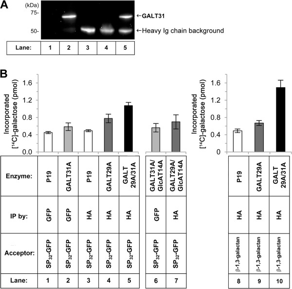
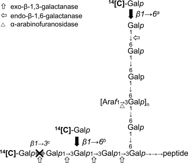
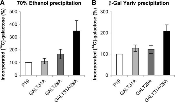
Results: We have identified a novel enzyme in the biosynthesis of the glycan moiety of arabinogalactan proteins. The At1g08280 (AtGALT29A) from Arabidopsis thaliana encodes a putative glycosyltransferase (GT), which belongs to the Carbohydrate Active Enzyme family GT29. AtGALT29A co-expresses with other arabinogalactan GTs, AtGALT31A and AtGLCAT14A. The recombinant AtGALT29A expressed in Nicotiana benthamiana demonstrated a galactosyltransferase activity, transferring galactose from UDP-galactose to a mixture of various oligosaccharides derived from arabinogalactan proteins. The galactose-incorporated products were analyzed using structure-specific hydrolases indicating that the recombinant AtGALT29A possesses β-1,6-galactosyltransferase activity, elongating β-1,6-galactan side chains and forming 6-Gal branches on the β-1,3-galactan main chain of arabinogalactan proteins. The fluorescence tagged AtGALT29A expressed in N. benthamiana was localized to Golgi stacks where it interacted with AtGALT31A as indicated by Förster resonance energy transfer. Biochemically, the enzyme complex containing AtGALT31A and AtGALT29A could be co-immunoprecipitated and the isolated protein complex exhibited increased level of β-1,6-galactosyltransferase activities compared to AtGALT29A alone.

Conclusions: AtGALT29A is a β-1,6-galactosyltransferase and can interact with AtGALT31A. The complex can work cooperatively to enhance the activities of adding galactose residues 6-linked to β-1,6-galactan and to β-1,3-galactan. The results provide new knowledge of the glycosylation process of arabinogalactan proteins and the functional significance of protein-protein interactions among O-glycosylation enzymes.


Figures

Figure 1
Figure 1
Identification of donor substrate for recombinant AtGALT29A. Affinity purified AtGALT29A (■) or P19 (□) was incubated with A: NDP-[14C]-sugars: UDP-[14C]-Xyl, UDP-[14C]-Glc, GDP-[14C]-Man and GDP-[14C]-Fuc (as MixI), and UDP-[14C]-GlcNAc, UDP-[14C]-GlcA and UDP-[14C]-Gal (as MixII); B: or individual NDP-[14C]-sugars from MixII using GAGP8 as acceptor substrate. Error bars showed standard deviations from n = 4. The result indicates that UDP-[14C]-Gal serves substrate for AtGALT29A.
Figure 2
Figure 2
Subcellular localization of AtGALT29A-mCer3 in N. benthamiana leaves. A-B: Confocal images of AtGALT29A-mCer3, STtmd-YFP (a Golgi marker) co-expressed transiently in N. benthamiana leaves. C: The overlay image of (A) and (B). The result indicates co-localization of ATGALT29A-mCer3 and STtmd-YFP in the Golgi apparatus. Scale bar = 5 μm.
Figure 3
Figure 3
Localization and FRET analysis for AtGALT29A and AtGALT31A. A-B: Confocal images of AtGALT31A-mCer3 and AtGALT29A-YFP co-expressed in N. benthamiana leaves. C: The overlay image of (A) and (B). AtGALT31A-mCer3 and AtGALT29A-YFP are co-localized in high frequency. D-G: Distribution histogram for pixel by pixel analysis of FRET [26]. FRET efficiency is expressed as FRET=, for example, FRET = 0.19 in (D) means that FRET efficiency is 19%; SEM, standard error of means; cell = number of cells analyzed. Scale bar = 5 μm.
Figure 4
Figure 4
Galactosyltransferase activity using the purified AtGALT29A/AtGALT31A complex in vitro. Microsomes were prepared from N. benthamiana leaves after expression of P19 only, AtGALT31A-GFP, HA-AtGALT29A or co-expression of HA-AtGALT29A and AtGALT31A-GFP, and subjected to immunoprecipitation using anti-GFP- or anti-HA-antibody. The conditions are indicated in the table at the bottom of (B). The immunoprecipitated samples were analyzed by the Western blot (A) and by the enzyme activity (B). A: The Western blot of P19, AtGALT31A-GFP, HA-AtGALT29A and AtGALT29A/AtGALT31A immunoprecipitated using GFP antibody. The result indicates co-purification of AtGALT31A-GFP (lane 5, indicated by the arrow at ca. 70 kDa) by immunoprecipitation of HA-AtGALT29A using anti-HA-antibody-agarose. The 50 kDa band detected in the lanes 3-5 is the heavy chain of HA antibody used for the immunoprecipitation, which is recognized by the secondary antibody used in the Western blot. B: Galactosyltransferase activity towards SP32-GFP and β-1,3-galactan acceptors. Affinity purified materials from the expression of P19 only, AtGALT31A-GFP, HA-AtGALT29A, or co-expression of HA-AtGALT29A and AtGALT31A-GFP using anti-GFP- or anti-HA-antibody were tested for enzyme activity using UDP-14[C]-Gal as substrate and SP32-GFP (lanes 1-5) or β-1,3-galactan (lanes 8-10) as acceptor, (n = 4). Control samples after co-expression of AtGALT31A-GFP or HA-AtGALT29A with HA-AtGLCAT14A (lane 6 and 7) were immunoprecipitated in the same way as for other samples and tested for the enzyme activity using UDP-14[C]-Gal as substrate and SP32-GFP as acceptor (lanes 6-7), (n = 3). These combinations are not suggested to form protein complexes based on the FRET analysis. Error bars showed standard deviations.
Figure 5
Figure 5
Simplified model structure of arabinogalactan and reaction sites of enzymes. The cleavage sites of the hydrolases (exo-β-1,3-galactanase, endo-β-1,6-galactanase, α-arabinofuranosidase) used in this paper are indicated. Recombinant AtGALT29A produced Gal incorporated products susceptible to the treatment of endo-β-1,6- and exo-β-1,3-galactanases (Figure 6), therefore three possible sites (β1 → 6a, b and β1 → 3c) are conceivable as the candidate sites of reaction. Towards β-1,3-galactan acceptor, both β1 → 6b and β1 → 3c galactosyltransferase activities are possible, but the main compound released by the exo-β-1,3-galactanase treatment was galactobiose, and not galactose (inset TLC in Figure 6C, D), indicating a β1 → 6b activity rather than β1 → 3c activity. Together with the β1 → 6a activity indicated by the endo-β-1,6-galactanase treatment, it is concluded that, AtGALT29A possesses β-1,6-galactosyltransferase activities both on β-1,3- and β-1,6-galactan (β1 → 6a, b activities).
Figure 6
Figure 6
Analysis of the sites of Gal incorporation in the products produced by AtGALT29A alone or the AtGALT29A/AtGALT31A complex. The [14C]-Gal incorporated products onto SP32-GFP (A, B, C) or onto β-1,3-galactan (D) from P19 [∙∙∙], HA-AtGALT29A [---], or co-immunoprecipitated HA-AtGALT29A/AtGALT31A-GFP complex [▬] were treated with A: endo-β-1,6-galactanase, B: endo-β-1,6-galactanase + α-arabinofuranosidase, C: exo-β-1,3-galactanase, or D: exo-β-1,3-galactanase, and separated by size exclusion chromatography using Superdex Peptide HR 10/30. The [14C]-Gal present in each fraction was evaluated by scintillation counting. Endo-β-1,6-galactanase, α-arabinofuranosidase, and exo-β-1,3-galactanase used in this study cleave β-1,6-linked unsubstituted galactotriose, terminal α-linked arabinofuranose, and β-1,3-linked galactooligosaccharides regardless the presence or absence of substitutions, respectively. Release of small [14C]-oligosaccharides by endo-β-1,6-galactanase indicates the [14C]-Gal incorporation to a part of β-1,6-galactotriose, while exo-β-1,3-galactanase releases [14C]-Gal monomer from β-1,3-linked galactan and [14C]-oligosaccharide (s) from side chains attached to β-1,3-linked galactan. From the [14C]-products made onto SP32-GFP and β-1,3-galactan, exo-β-1,3-galactanase released mainly [14C]-galactobiose analyzed by TLC (inset C and D), indicating the incorporation of single [14C]-Gal to β-1,3-linked Gal at O6 in the [14C]-products. From any treatments (A-D), higher amount of small [14C]-oligosaccharides are released from the [14C]-products made by AtGALT29A/AtGALT31A complex compared to that from a single enzyme. The results indicate that AtGALT29A possesses β-1,6-GalT activities elongating β-1,6-galactan and forming 6-Gal branches on β-1,3-galatan, and the β-1,6-GalT activities are increased when AtGALT29A is in a protein complex with AtGALT31A.
Figure 7
Figure 7
Galactosyltransferase activity in intact microsomes isolated from N. benthamiana after co-expression of HA-AtGALT29A and AtGALT31A-GFP. Microsomes were incubated with exogenously added UDP-[14C]-Gal and the [14C]-Gal incorporation to luminal endogenous materials were analyzed by precipitation either by A: 70% ethanol or B: β-Gal Yariv reagent. Error bars showed standard deviations from n = 4.

References

    1. Seifert GJ, Roberts K. Annual Review of Plant Biology, Volume 58. Palo Alto: Annual Reviews; 2007. The biology of arabinogalactan proteins; pp. 137–161. - PubMed
    1. Pennell RI, Janniche L, Kjellbom P, Scofield GN, Peart JM, Roberts K. Developmental Regulation of a Plasma-Membrane Arabinogalactan Protein Epitope in Oilseed Rape Flowers. Plant Cell. 1991;3(12):1317–1326. - PMC - PubMed
    1. Pennell RI, Knox JP, Scofield GN, Selvendran RR, Roberts K. A Family of Abundant Plasma-Membrane Associated Glycoproteins Related to the Arabinogalactan Proteins Is Unique to Flowering Plants. J Cell Biol. 1989;108(5):1967–1977. - PMC - PubMed
    1. Stacey NJ, Roberts K, Carpita NC, Wells B, McCann MC. Dynamic changes in cell surface molecules are very early events in the differentiation of mesophyll cells from Zinnia elegans into tracheary elements. Plant J. 1995;8(6):891–906.
    1. Stacey NJ, Roberts K, Knox JP. Patterns of Expression of the Jim4 Arabinogalactan-Protein Epitope in Cell-Cultures and during Somatic Embryogenesis in Daucus-Carota L. Planta. 1990;180(2):285–292. - PubMed

Publication types

MeSH terms