. 2014 Jun;155(6):2301-13.
doi: 10.1210/en.2013-1933. Epub 2014 Apr 2.

Epidermal growth factor receptor (EGFR) signaling is a key mediator of hormone-induced leukocyte infiltration in the pubertal female mammary gland

Affiliations

Epidermal growth factor receptor (EGFR) signaling is a key mediator of hormone-induced leukocyte infiltration in the pubertal female mammary gland

Mark D Aupperlee et al. Endocrinology. 2014 Jun.

Abstract

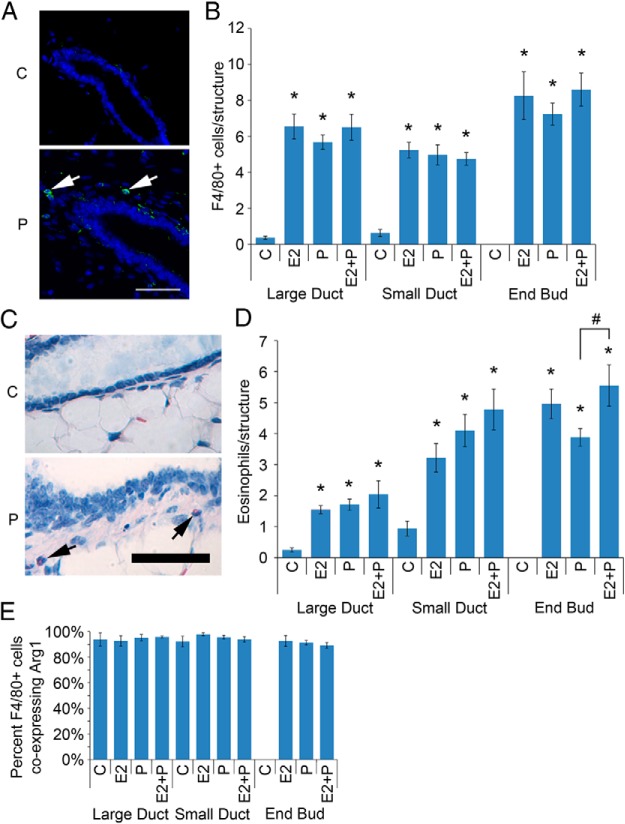
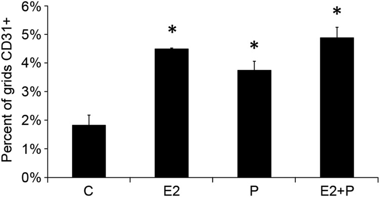
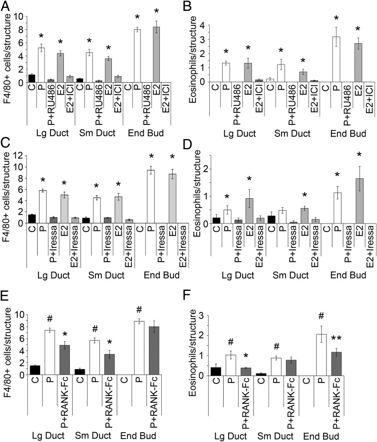
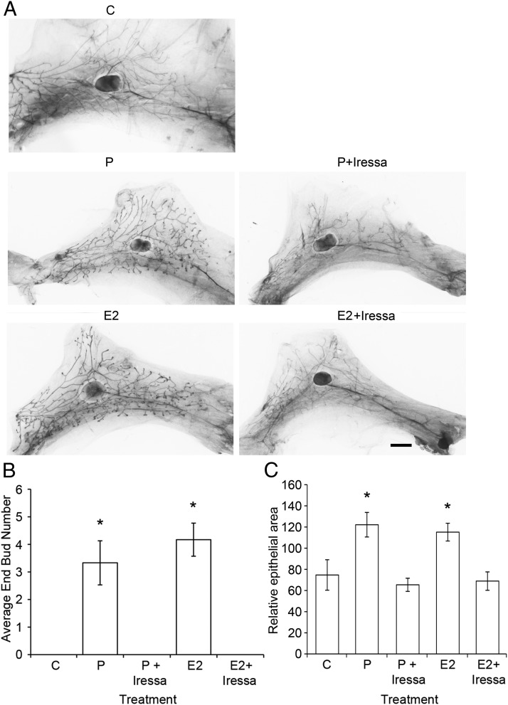
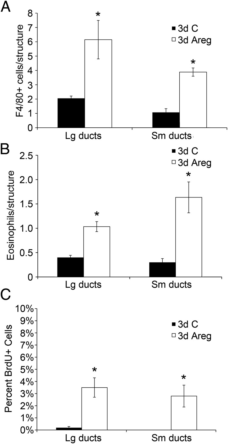
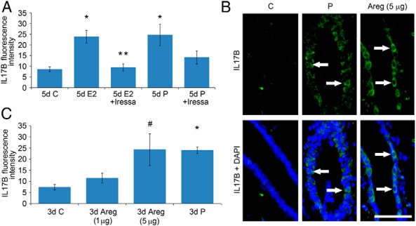
It is well documented that macrophages and eosinophils play important roles in normal murine pubertal mammary gland development. Although it is accepted that estrogen (E) and progesterone (P) are key players in mammary gland development, the roles these hormones might play in regulating the actions of leukocytes in that process is an understudied area. We show here that P and E, respectively, induce unique, but overlapping, sets of proinflammatory and angiogenic cytokines and chemokines, in the pubertal female BALB/c mammary gland, as well as induce infiltration of macrophages and eosinophils to the mammary periepithelium. This extends earlier studies showing P induction of proinflammatory products in pubertal and adult mammary epithelial organoids and P-induced in vivo infiltration of leukocytes to the adult mammary periepithelium. Importantly, epidermal growth factor receptor-signaling, which is likely mediated by amphiregulin (Areg), a downstream mediator of E and P, is both necessary and sufficient for both E- and P-induced recruitment of macrophages and eosinophils to the pubertal mammary periepithelium. We further show that receptor activator of nuclear factor κB ligand (RANKL), although not sufficient of itself to cause macrophage and eosinophil recruitment, contributes to an optimal response to P. The potency of Areg is highlighted by the fact that it is sufficient to induce macrophage and eosinophil recruitment at levels equivalent to that induced by either E or P. Our finding of a dominant role for Areg in hormonally induced leukocyte recruitment to the pubertal mammary gland parallels its dominance in regulating ductal outgrowth and its role in P-induced proliferation in the pubertal gland.


Figures

Figure 1.
Figure 1.
Both E2 and P regulate macrophage and eosinophil recruitment to the pubertal mammary gland. Pubertal 4-week-old BALB/c mice were OVX, allowed to recover for 3 weeks, and then treated for 5 days with vehicle control (C), E2, P, or E2+P. A, Immunofluorescent detection of F4/80 (green) in C and P-treated mammary gland. Nuclei were counterstained with DAPI (blue). Scale bar, 25 μm. B, F4/80-positive cell recruitment to the periepithelium was analyzed for large ducts, small ducts, and duct ends. The values represent the mean ± SEM (n = 6 animals per treatment). The number of macrophages recruited to periepithelial structures upon treatment with E2, P, and E2+P was above C levels (*, P < .05). C, Histochemical detection of eosinophils (arrows) in C and P-treated mammary gland. Nuclei were counterstained with hematoxylin (blue). Scale bar, 50 μm. D, Eosinophil recruitment to the periepithelium was analyzed for large ducts, small ducts, and duct ends. The number of eosinophils recruited to periepithelial structures upon treatment with E2, P, and E2+P was above C levels (*, P < .05). The number of eosinophils recruited to the periepithelium of P-treated EBs was reduced compared with E2+P-treated EBs (#, P < .05). The values represent the mean ± SEM (n = 6 animals per treatment). E, Macrophage analysis by dual immunofluorescent detection of F4/80 and Arg1. The percentage of F4/80-positive cells coexpressing Arg1 is presented. The values represent the mean ± SEM (n = 3 animals per treatment).
Figure 2.
Figure 2.
E2 and P increase angiogenesis in the pubertal mammary gland. Pubertal 4-week-old BALB/c mice were OVX, allowed to recover for 3 weeks, and then treated for 5 days with vehicle control (C), E2, P, or E2+P. Blood vessel density was measured, as described in Materials and Methods, by the area occupied by CD31-positive vessels near mammary epithelium. The values represent the mean ± SEM (n = 3 animals per treatment). CD31 staining was greater in E2-, P-, and E2+P-treated mammary glands (*, P < .05) compared with Cs.
Figure 3.
Figure 3.
Areg and RANKL are mediators of hormone-induced macrophage and eosinophil recruitment to mammary epithelium. Pubertal 4-week-old BALB/c mice were OVX, allowed to recover for 3 weeks, and then treated for 5 days with vehicle control (C), E2, or P. A and B, Both macrophage (A) and eosinophil (B) recruitment to mammary gland epithelial structures by E2 and P were inhibited by E2+ICI and P+RU486. The numbers of macrophages and eosinophils recruited to periepithelial structures upon treatment with E2 or P were above that of other treatments (*, P < .05). C and D, Both macrophage (C) and eosinophil (D) recruitment to mammary gland epithelial structures by E2 and P were inhibited by E2+Iressa and P+Iressa. E2- and P-induced macrophage recruitment was completely inhibited in large and small ducts by the addition of Iressa. No EBs were detected in Iressa-treated mammary glands. The numbers of macrophages or eosinophils recruited to periepithelial structures upon treatment with E2 or P were above that of other treatments (*, P < .05). E and F, Both macrophage (E) and eosinophil (F) recruitment to mammary gland epithelial structures by P were inhibited by P+RANK-Fc. The numbers of macrophages or eosinophils recruited to periepithelial structures upon treatment P were above that of C treatments (#, P < .05). P+RANK-Fc decreased macrophage recruitment to large and small ducts compared with P alone (*, P < .05). P+RANK-Fc decreased eosinophil recruitment to large ducts (*, P < .05) and EBs (**, P < .1) compared with P alone. A–F, The values represent the mean ± SEM (n = 3 animals per treatment).
Figure 4.
Figure 4.
Iressa inhibits both E2- and P-induced EB formation and epithelial outgrowth. Pubertal 4-week-old BALB/c were OVX, allowed to recover for 3 weeks, and then treated for 5 days with vehicle control (C), E2 ± Iressa (300 mg/kg), or P ± Iressa. A, Whole mounts showing effect of treatment on EB formation after 5 days of C, E2, E2+Iressa, P, or P+Iressa. Note the absence of EBs in Iressa-treated mammary glands. Scale bar, 2 mm. B, EB quantitation from C and treated whole mounts. Values represent the mean ± SEM number of EBs in an inguinal mammary gland (n = 4 mice). E2 and P increased the average EB number (*, P < .05), and this increase was inhibited by the addition of Iressa. C, Epithelial area quantitation from C and treated whole mounts. Values represent the mean ± SEM epithelial area in a pair of inguinal mammary glands (n = 4 mice). E2 and P increased the average epithelial area (*, P < .05), and this increase was inhibited by the addition of Iressa.
Figure 5.
Figure 5.
Areg increased macrophage and eosinophil recruitment and mammary epithelial cell proliferation. Pubertal 4-week-old BALB/c mice were OVX, allowed to recover for 3 weeks, and then treated for 3 days with bovine serum albumin (BSA) control (C) or Areg. A and B, Both macrophage (A) and eosinophil recruitment (B) were significantly increased by Areg over BSA Cs across all mammary epithelial structures (*, P < .05). C, The percentage of BrdU+ cells was increased by Areg over BSA Cs across all mammary epithelial structures (*, P < .05). A–C, The values represent the mean ± SEM (n = 3 animals per treatment).
Figure 6.
Figure 6.
IL-17B is regulated by E2 and P through Areg. A, Pubertal 4-week-old BALB/c mice were OVX, allowed to recover for 3 weeks, and then treated for 5 days with vehicle control (C), E2, E2+Iressa, P, or P+Iressa. A, IL-17B was measured in mammary epithelium by immunofluorescence, as intensity above background. The values represent the mean ± SEM (n = 3 animals per treatment). Five-day E2 and P treatment increased IL-17B compared with C (*, P < .05). E2+Iressa inhibited E2-induced increases in IL-17B (**, P < .05). B and C, Pubertal 4-week-old BALB/c mice were OVX, allowed to recover for 3 weeks, and then treated for 3 days with BSA C, 1 or 3 μg of Areg, or P. B, Immunofluorescent detection of IL-17B (green). White arrows mark examples of cytoplasmic IL-17B expression colocalizing to DAPI staining in mammary epithelial cells. Scale bar, 25 μm. C, IL-17B was measured in mammary epithelium by immunofluorescence, as intensity above background. The values represent the mean ± SEM (n = 3 animals per treatment). Three-day P treatment (*, P < .05) and 3-day 5-μg Areg treatment (#, P < .05) increased IL-17B compared with C treatment.

References

    1. Gouon-Evans V, Rothenberg ME, Pollard JW. Postnatal mammary gland development requires macrophages and eosinophils. Development. 2000;127:2269–2282 - PubMed
    1. Lilla JN, Werb Z. Mast cells contribute to the stromal microenvironment in mammary gland branching morphogenesis. Dev Biol. 2010;337:124–133 - PMC - PubMed
    1. Haslam SZ, Drolet A, Smith K, Tan M, Aupperlee M. Progestin-regulated luminal cell and myoepithelial cell-specific responses in mammary organoid culture. Endocrinology. 2008;149:2098–2107 - PMC - PubMed
    1. Santos SJ, Aupperlee MD, Xie J, et al. Progesterone receptor A-regulated gene expression in mammary organoid cultures. J Steroid Biochem Mol Biol. 2009;115:161–172 - PMC - PubMed
    1. Aupperlee MD, Drolet AA, Durairaj S, Wang W, Schwartz RC, Haslam SZ. Strain-specific differences in the mechanisms of progesterone regulation of murine mammary gland development. Endocrinology. 2009;150:1485–1494 - PMC - PubMed

Publication types