

A comparison of the diagnostic accuracy of MARS MRI and ultrasound of the painful metal-on-metal hip arthroplasty
- PMID: 24694273
- PMCID: PMC4105768
- DOI: 10.3109/17453674.2014.908345
A comparison of the diagnostic accuracy of MARS MRI and ultrasound of the painful metal-on-metal hip arthroplasty
Abstract
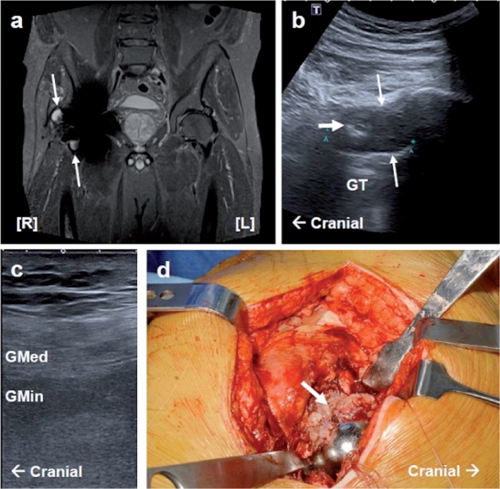
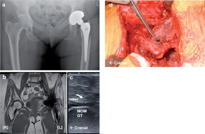
Background and purpose: Metal artifact reduction sequence (MARS) MRI and ultrasound scanning (USS) can both be used to detect pseudotumors, abductor muscle atrophy, and tendinous pathology in patients with painful metal-on-metal (MOM) hip arthroplasty. We wanted to determine the diagnostic test characteristics of USS using MARS MRI as a reference for detection of pseudotumors and muscle atrophy. PatienTS AND METHODS: We performed a prospective cohort study to compare MARS MRI and USS findings in 19 consecutive patients with unilateral MOM hips. Protocolized USS was performed by consultant musculoskeletal radiologists who were blinded regarding clinical details. Reports were independently compared with MARS MRI, the imaging gold standard, to calculate predictive values.
Results: The prevalence of pseudotumors on MARS MRI was 68% (95% CI: 43-87) and on USS it was 53% (CI: 29-76). The sensitivity of USS in detecting pseudotumors was 69% (CI 39-91) and the specificity was 83% (CI: 36-97). The sensitivity of detection of abductor muscle atrophy was 47% (CI: 24-71). In addition, joint effusion was detected in 10 cases by USS and none were seen by MARS MRI.
Interpretation: We found a poor agreement between USS and MARS MRI. USS was inferior to MARS MRI for detection of pseudotumors and muscle atrophy, but it was superior for detection of joint effusion and tendinous pathologies. MARS MRI is more advantageous than USS for practical reasons, including preoperative planning and longitudinal comparison.
Figures
References
-
- Almousa SA, Greidanus NV, Masri BA, Duncan CP, Garbuz DS. The Natural History of Inflammatory Pseudotumors in asymptomatic patient... . Clin Orthop. 2013;471(12):3814–21. - PMC - PubMed
-
- Bal BS, Lowe JA. Muscle damage in minimally invasive total hip arthroplasty: MRI evidence... . Instr Course Lect. 2008;57:223–9. - PubMed
-
- Davies AP, Willert HG, Campbell PA, Learmonth ID, Case CP. An unusual lymphocytic perivascular infiltration in tissues around conte... . J Bone Joint Surg (Am) 2005;87(1):18–27. - PubMed
-
- Douis H, Dunlop DJ, Pearson AM, O’Hara JN, James SL. The role of ultrasound in the assessment of post-operative complications... . Skeletal Radiol. 2012;41:9, 1035–46. - PubMed
-
- Fang CS, Harvie P, Gibbons CL, Whitwell D, Athanasou N A, Ostlere S. The imaging spectrum of peri-articular inflammatory masses following met... . Skeletal Radiol. 2008;37:8, 715–22. - PubMed
Publication types
MeSH terms
Substances
Grants and funding
LinkOut - more resources
Full Text Sources
Other Literature Sources
Medical
Miscellaneous