. 2014 Jun;25(11):1715-29.
doi: 10.1091/mbc.E13-12-0730. Epub 2014 Apr 2.

Primary cilia in stem cells and neural progenitors are regulated by neutral sphingomyelinase 2 and ceramide

Affiliations

Primary cilia in stem cells and neural progenitors are regulated by neutral sphingomyelinase 2 and ceramide

Qian He et al. Mol Biol Cell. 2014 Jun.

Abstract

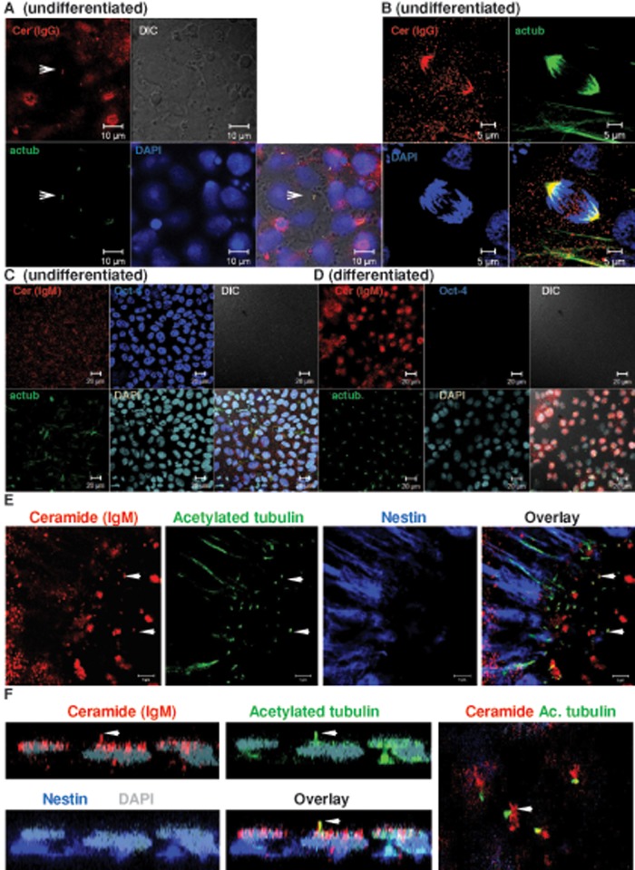
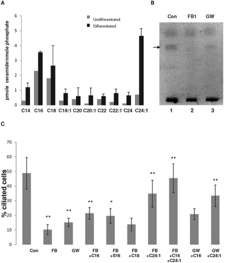
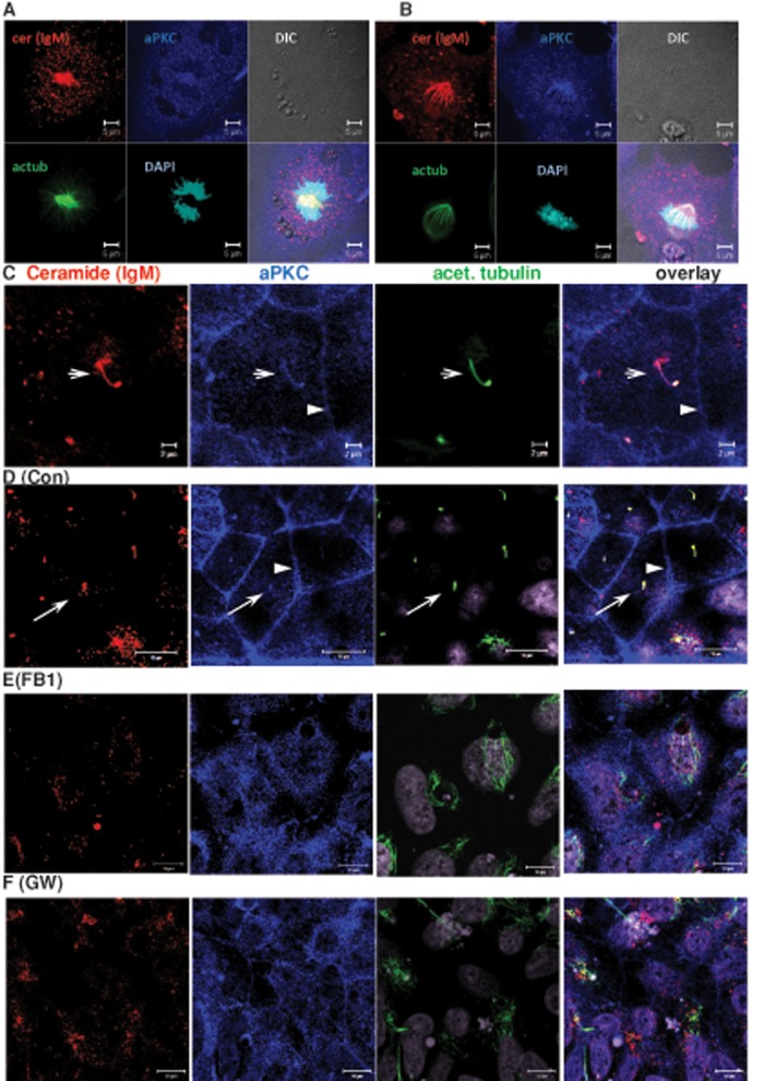
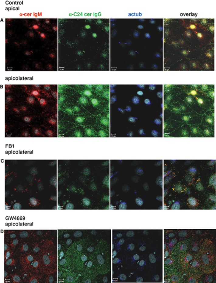
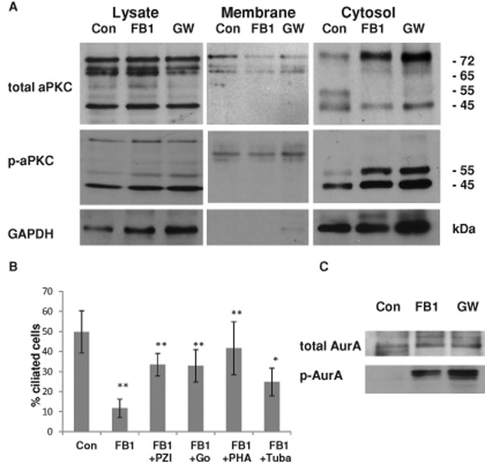
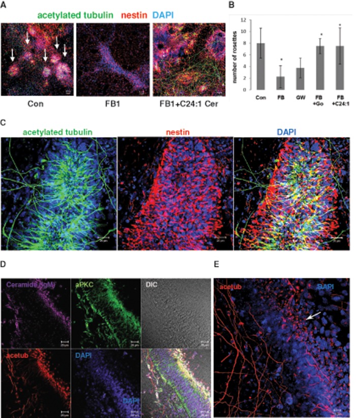
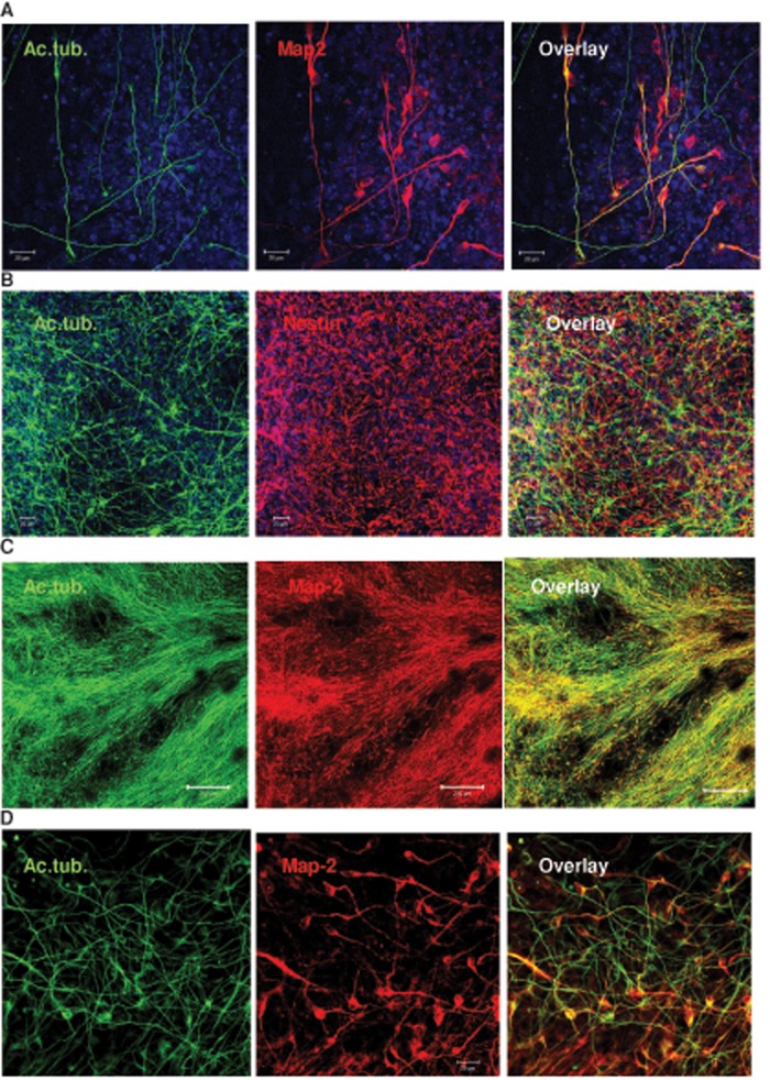
We show here that human embryonic stem (ES) and induced pluripotent stem cell-derived neuroprogenitors (NPs) develop primary cilia. Ciliogenesis depends on the sphingolipid ceramide and its interaction with atypical PKC (aPKC), both of which distribute to the primary cilium and the apicolateral cell membrane in NP rosettes. Neural differentiation of human ES cells to NPs is concurrent with a threefold elevation of ceramide-in particular, saturated, long-chain C16:0 ceramide (N-palmitoyl sphingosine) and nonsaturated, very long chain C24:1 ceramide (N-nervonoyl sphingosine). Decreasing ceramide levels by inhibiting ceramide synthase or neutral sphingomyelinase 2 leads to translocation of membrane-bound aPKC to the cytosol, concurrent with its activation and the phosphorylation of its substrate Aurora kinase A (AurA). Inhibition of aPKC, AurA, or a downstream target of AurA, HDAC6, restores ciliogenesis in ceramide-depleted cells. Of importance, addition of exogenous C24:1 ceramide reestablishes membrane association of aPKC, restores primary cilia, and accelerates neural process formation. Taken together, these results suggest that ceramide prevents activation of HDAC6 by cytosolic aPKC and AurA, which promotes acetylation of tubulin in primary cilia and, potentially, neural processes. This is the first report on the critical role of ceramide generated by nSMase2 in stem cell ciliogenesis and differentiation.


Figures

FIGURE 1:
FIGURE 1:
Ceramide is colocalized with acetylated tubulin in primary cilia and the mitotic spindle. Human ES cells were cultivated feeder free and then subjected to immunocytochemistry using antibodies against ceramide (rabbit IgG or mouse IgM, red) and acetylated tubulin (green). (A) Interphase; arrow points at primary cilium. Bar, 10 μm. (B) Metaphase. Bar, 5 μm. (C, D) Neural differentiation suppresses expression of Oct-4 concurrent with elevated expression of ceramide and formation of primary cilia (C is before and D is after neural differentiation). Bar, 20 μm. (E) Primary cilia are formed in nestin-expressing NPs in neural rosettes. In NPs, ceramide is colocalized with primary cilia and vesicles at the cilium bases (arrows). Bar, 5 μm. (F) A z-scan of individual cell in neural rosette. Arrow indicates colocalization of ceramide with acetylated tubulin in primary cilium. Right, apical plane of area used for z-scan.
FIGURE 2:
FIGURE 2:
Ceramide depletion reduces ciliogenesis, whereas addition of ceramide rescues cilium formation. Mass spectrometric analysis of ceramide species in undifferentiated human ES cells and ES cell-derived NPs (A) and HPTLC analysis (B) of ceramide in NPs in the presence or absence of inhibitors for ceramide generation (FB1 or GW4869). Arrow in B points at ceramide band stained with cupric acetate/charring. (C) Proportion of ciliated cells (length of cilia >1 μm) in NPs with reduced ceramide levels and after the addition of exogenous ceramide (C16, C16:0 ceramide; C18, C18:0 ceramide; C24:1, C24:1 ceramide) or ceramide analogues (S16, N-palmitoyl serinol). N > 5; p value for difference between controls and FB1- or GW4869-treated cells, **p < 0.01, and for difference between inhibitor-treated cells and those with C16:0 ceramide, S16, or C24:1 ceramide exogenously added, *p < 0.05, **p < 0.01. There is no significant difference between FB1- or GW4869-treated cells and those given exogenous C18:0 ceramide.
FIGURE 3:
FIGURE 3:
Ceramide depletion leads to translocation of aPKC from the primary cilium and the apicolateral cell membrane to the cytosol. NPs were analyzed using immunocytochemistry with anti-ceramide mouse IgM (red) and antibodies against acetylated tubulin (mouse IgG, green) and aPKC (C20, rabbit IgG, blue). (A, B) Mitotic cells. Note colocalization of ceramide with mitotic spindle. Bar, 5 μm. (C) Interphase (arrow points at primary cilium). 4′,6-Diamidino-2-phenylindole (DAPI) staining is pseudocolored in gray. Bar, 2 μm. (D–F) NPs differentiated from control (D) and FB1- (E) and GW4869-treated (F) human ES cells, followed by immunocytochemistry using antibodies as described for (A–C). aPKC is colocalized with the primary cilium (arrow) and the apicolateral cell membrane (arrowhead) of control cells (D). Colocalization is lost when cells are depleted of ceramide by treatment with FB1 (E) or GW4869 (F). Bars, 10 μm.
FIGURE 4:
FIGURE 4:
C16:0 ceramide is localized at primary cilia, whereas C24:0/24:1 ceramide is at primary cilia and the apicolateral cell membrane. Neural differentiation was performed in the presence or absence of FB1 or GW4869, followed by immunocytochemistry using antibodies against C16:0 ceramide (anti-cerIgM) or C24:0/C24:1 ceramide (anti-C24). DAPI staining is pseudocolored in gray, anti-ceramide IgM in red, anti-C24 ceramide IgG in green, and acetylated tubulin in blue. (A) Control, apical plane (arrow points at colocalization of anti-cerIgM, anti-C24, and anti-acetylated tubulin antibodies at primary cilia). (B) Control, apicolateral plane (arrow points at apicolateral cell membrane). (C) FB1-treated cells. (D) GW4869-treated cells. Bars, 10 μm.
FIGURE 5:
FIGURE 5:
In ceramide-depleted cells, activation of cytosolic aPKC reduces ciliogenesis, whereas inhibition of HDAC6 and AurA rescues cilia. (A) Control and FB1- or GW4869-treated NPs were subjected to subcellular fractionation using differential centrifugation. The cytosolic fraction of ceramide-depleted cells contains a phosphorylated, 55-kDa isoform of aPKC (aPKCcat). In addition, there are 65- and 45-kDa isoforms, the latter of which is phosphorylated but it is also present in control cells. (B) Inhibition of aPKC with pseudo substrate inhibitor peptide of aPKC (PZI) or Go6983 (Go), or AurA and HDAC6 inhibition with PHA-680632 (PHA) and tubacin (Tuba), can partially rescue ciliogenesis in FB1-treated NPs. N ≥ 5, *p < 0.05 or **p < 0.01 for comparison of treated cells with control. (C) Ceramide depletion leads to enhanced phosphorylation of AurA in the cytosolic fraction.
FIGURE 6:
FIGURE 6:
C24:1 ceramide restores NP rosettes in ceramide-depleted cells. Human ES cells were differentiated for 5 d in the presence of 20 μM FB1 with or without 2 μM C24:1 ceramide, followed by immunocytochemistry for acetylated tubulin (mouse or rabbit IgG), aPKC (rabbit IgG), ceramide (mouse IgM), or nestin (mouse IgG). A. Formation of neural rosettes (arrows) is severely reduced in ceramide-depleted NPs. (B) Treatment with C24:1 ceramide restores the number of NP rosettes (>100 μm diameter) similar to the effect of aPKC inhibition with Go6983. N = 3, p < 0.05 for comparison of FB1-treated and C24:1 ceramide–treated cells. (C–E) C24:1 ceramide treatment promotes formation of acetylated tubulin–labeled processes and polarized neural tube–like structures (arrow in E points at primary cilia at apical side). Bars, 50 μm (A), 20 μm (C, D), and 10 μm (E).
FIGURE 7:
FIGURE 7:
C24:1 ceramide promotes neural process formation in human ES and iPS cells. Human ES or iPS cells were treated as described in the legend to Figure 6. (A) Colocalization of acetylated tubulin (green) and Map-2 (red) shows that many processes originate in neural progenitors or differentiated neurons. (B–D) Acetylated tubulin–labeled processes and nestin-labeled NPs are also visible in iPS cells that were differentiated following the protocol used for human ES cells. Neural processes of C24:1 ceramide–treated iPS cells are elongated (>200 μm) and colabeled for Map-2. Bars, 20 μm (A, B, D), 200 μm (C).

Similar articles

Cited by

References

    1. Amador-Arjona A, Elliott J, Miller A, Ginbey A, Pazour GJ, Enikolopov G, Roberts AJ, Terskikh AV. Primary cilia regulate proliferation of amplifying progenitors in adult hippocampus: implications for learning and memory. J Neurosci. 2011;31:9933–9944. - PMC - PubMed
    1. Aubin I, et al. A deletion in the gene encoding sphingomyelin phosphodiesterase 3 (Smpd3) results in osteogenesis and dentinogenesis imperfecta in the mouse. Nat Genet. 2005;37:803–805. - PubMed
    1. Bieberich E, Kawaguchi T, Yu RK. N-acylated serinol is a novel ceramide mimic inducing apoptosis in neuroblastoma cells. J Biol Chem. 2000;275:177–181. - PubMed
    1. Bieberich E, MacKinnon S, Silva J, Noggle S, Condie BG. Regulation of cell death in mitotic neural progenitor cells by asymmetric distribution of prostate apoptosis response 4 (PAR-4) and simultaneous elevation of endogenous ceramide. J Cell Biol. 2003;162:469–479. - PMC - PubMed
    1. Bougie JK, Cai D, Hastings M, Farah CA, Chen S, Fan X, McCamphill PK, Glanzman DL, Sossin WS. Serotonin-induced cleavage of the atypical protein kinase C Apl III in Aplysia. J Neurosci. 2012;32:14630–14640. - PMC - PubMed

Publication types

MeSH terms

LinkOut - more resources