. 2014 Apr 3:4:4577.
doi: 10.1038/srep04577.

FALCON: fast and unbiased reconstruction of high-density super-resolution microscopy data

Affiliations

FALCON: fast and unbiased reconstruction of high-density super-resolution microscopy data

Junhong Min et al. Sci Rep. .

Abstract

Super resolution microscopy such as STORM and (F)PALM is now a well known method for biological studies at the nanometer scale. However, conventional imaging schemes based on sparse activation of photo-switchable fluorescent probes have inherently slow temporal resolution which is a serious limitation when investigating live-cell dynamics. Here, we present an algorithm for high-density super-resolution microscopy which combines a sparsity-promoting formulation with a Taylor series approximation of the PSF. Our algorithm is designed to provide unbiased localization on continuous space and high recall rates for high-density imaging, and to have orders-of-magnitude shorter run times compared to previous high-density algorithms. We validated our algorithm on both simulated and experimental data, and demonstrated live-cell imaging with temporal resolution of 2.5 seconds by recovering fast ER dynamics.


Figures
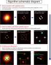
Figure 1
Figure 1. Schematic illustration for FALCON.
For a detailed description, see Supplementary note.
Figure 2
Figure 2. Taylor approximation of the shifted PSF for continuous refinement of FALCON.
(a) First-order Taylor approximation of the shifted PSF: the shifted PSF is approximated by the PSF centered on the sub-pixel and the derivative of the PSF. (b) Approximation error of Gaussian PSF of 360 nm FWHM. The error of the approximation does not exceed 2% up to 30 nm displacement.
Figure 3
Figure 3. Numerical analysis of the continuous refinement on 50,000 simulated images.
In each image, a single molecule is randomly placed with uniform probability distribution within a single sub-pixel area (a–c) or along a diagonal line (d–f). Histograms of ground-truth positions (a,d), histograms of the centroid fit that is applied to the high resolution images reconstructed by a sparsity-based deconvolution (l1 minimization) (b,e) and histograms of FALCON (c,f).
Figure 4
Figure 4. Performances of FALCON in comparison with Least-square fitting, DAOSTORM and CSSTORM over a wide range of imaging densities.
Simulation on the random distribution of molecules over a wide range of imaging densities with high-photon emission rates: recall rates (a), localization accuracy (b) and low-photon emission rates: recall rates (c), localization accuracy (d). The error bars represent standard deviations.
Figure 5
Figure 5. Performances of FALCON in comparison with DAOSTORM and CSSTORM over a wide range of PSF widths from 250 nm to 430 nm.
Simulated data with high-photon emission rates: (a) molecular recall rates, (b) localization accuracy and low-photon emission rates: (c) molecular recall rates, (d) localization accuracy. The error bars indicate standard deviations.
Figure 6
Figure 6. Performance analysis for synthetic “ring” phantoms with various radii.
(a) Rendered images of the algorithms overlaid by a cyan circle with 150 nm radius as a ground truth. (b) Histogram of the distances from localized molecules by FALCON, DAOSTORM, and CSSTORM to the center of the ring in (a). (c) The differences between the true radius and the distances on average along various radii from 150 nm to 400 nm with 4 fixed molecules on the circle in every simulated image. The error bars represent standard deviations.
Figure 7
Figure 7. FALCON performance on fixed microtubules data.
(a) FALCON reconstructed α-tubulin subunits of microtubules (MTs) labeled with Alexa 647 in Cos-7 cells; 500 raw frames were used to reconstruct a super-resolution image. White box highlights the intersection of MTs reconstructed by FALCON. (b) Fourier ring correlation(FRC) analysis on the reconstructed images by DAOSTORM, CSSTORM and FALCON. For the FRC analysis, fiducial beads indicated by yellow circles are discarded. The error bars represent standard deviations. Cross-sectional profiles across region in white box (yellow line) were measured by DAOSTORM, CSSTORM and FALCON, and plotted in (c). Scale bars are 1 μm in (a).
Figure 8
Figure 8. FALCON performance on live ER data.
Live imaging of the endoplasmic reticulum protein, reticulon-4 fused to tdEos imaged over 20 seconds in a U2OS cell. Conventional (a–e) and super-resolution (f–j) snapshots are shown with a 2.5 seconds temporal resolution. The average size of tubules measured from the reconstruction is approximately 60 nm (at FWHM). Yellow markers in (g) highlight a representative tubule width. The dynamic motions of these structures are highlighted in (g–j) indicated by white and blue arrows and dashed, yellow circles. All scale bars are 1 μm.

References

    1. Rust M. J., Bates M. & Zhuang X. Sub-diffraction-limit imaging by stochastic optical reconstruction microscopy STORM. Nat. Methods 3, 793–796 (2006). - PMC - PubMed
    1. Hess S. T., Girirajan T. P. & Mason M. D. Ultra-high resolution imaging by fluorescence photoactivation localization microscopy. Biophys. J. 91, 4258 (2006). - PMC - PubMed
    1. Betzig E. et al. Imaging intracellular fluorescent proteins at nanometer resolution. Science 313, 1642–1645 (2006). - PubMed
    1. Smith C. S., Joseph N., Rieger B. & Lidke K. A. Fast, single-molecule localization that achieves theoretically minimum uncertainty. Nat. Methods 7, 373–375 (2010). - PMC - PubMed
    1. Henriques R. et al. Quickpalm: 3d real-time photoactivation nanoscopy image processing in imagej. Nat. Methods 7, 339–340 (2010). - PubMed

Publication types