

Diversity of environmental single-stranded DNA phages revealed by PCR amplification of the partial major capsid protein
- PMID: 24694711
- PMCID: PMC4184009
- DOI: 10.1038/ismej.2014.43
Diversity of environmental single-stranded DNA phages revealed by PCR amplification of the partial major capsid protein
Abstract
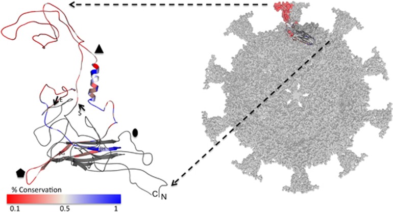
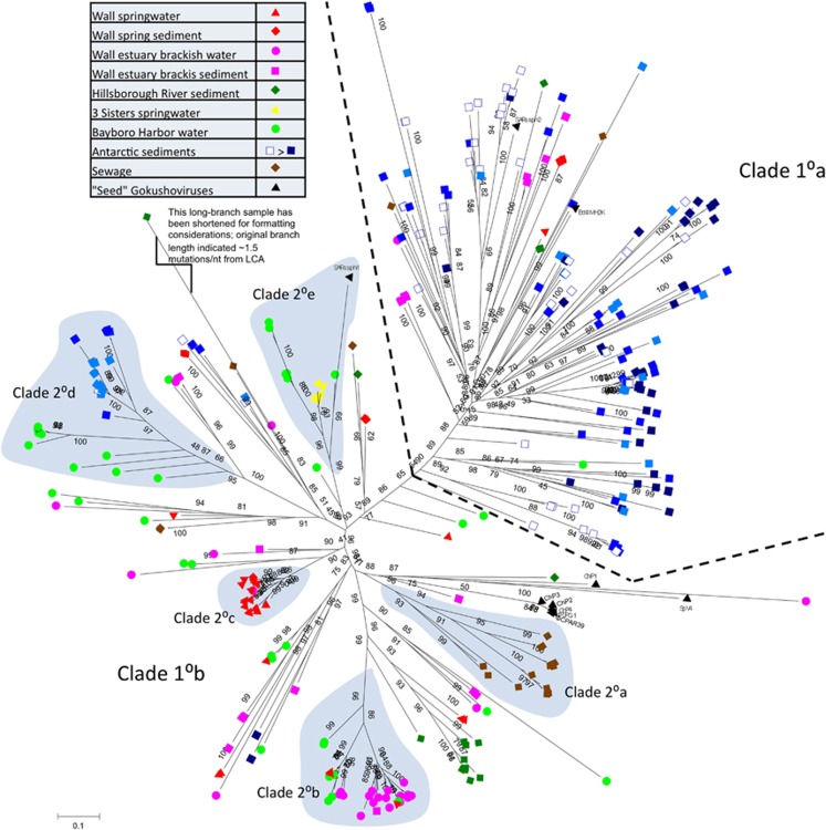
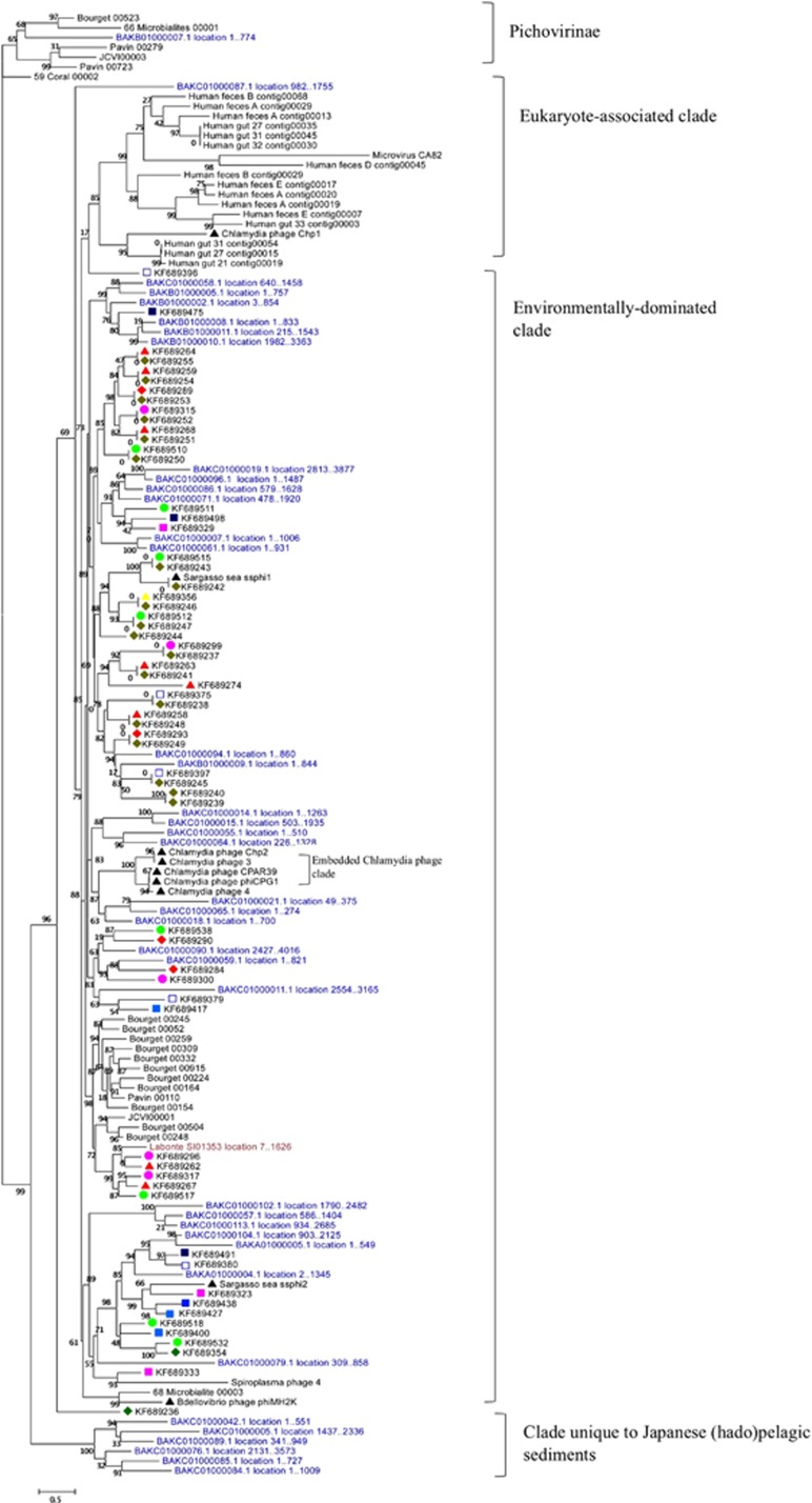
The small single-stranded DNA (ssDNA) bacteriophages of the subfamily Gokushovirinae were traditionally perceived as narrowly targeted, niche-specific viruses infecting obligate parasitic bacteria, such as Chlamydia. The advent of metagenomics revealed gokushoviruses to be widespread in global environmental samples. This study expands knowledge of gokushovirus diversity in the environment by developing a degenerate PCR assay to amplify a portion of the major capsid protein (MCP) gene of gokushoviruses. Over 500 amplicons were sequenced from 10 environmental samples (sediments, sewage, seawater and freshwater), revealing the ubiquity and high diversity of this understudied phage group. Residue-level conservation data generated from multiple alignments was combined with a predicted 3D structure, revealing a tendency for structurally internal residues to be more highly conserved than surface-presenting protein-protein or viral-host interaction domains. Aggregating this data set into a phylogenetic framework, many gokushovirus MCP clades contained samples from multiple environments, although distinct clades dominated the different samples. Antarctic sediment samples contained the most diverse gokushovirus communities, whereas freshwater springs from Florida were the least diverse. Whether the observed diversity is being driven by environmental factors or host-binding interactions remains an open question. The high environmental diversity of this previously overlooked ssDNA viral group necessitates further research elucidating their natural hosts and exploring their ecological roles.
Figures
Similar articles
-
Adaptation of the polony technique to quantify Gokushovirinae, a diverse group of single-stranded DNA phage.Environ Microbiol. 2021 Nov;23(11):6622-6636. doi: 10.1111/1462-2920.15805. Epub 2021 Oct 18. Environ Microbiol. 2021. PMID: 34623742
-
Structure and diversity of ssDNA Microviridae viruses in two peri-alpine lakes (Annecy and Bourget, France).Res Microbiol. 2015 Oct;166(8):644-54. doi: 10.1016/j.resmic.2015.07.003. Epub 2015 Jul 27. Res Microbiol. 2015. PMID: 26226335
-
Unprecedented Diversity of ssDNA Phages from the Family Microviridae Detected within the Gut of a Protochordate Model Organism (Ciona robusta).Viruses. 2018 Jul 31;10(8):404. doi: 10.3390/v10080404. Viruses. 2018. PMID: 30065169 Free PMC article.
-
Single-stranded DNA phages: from early molecular biology tools to recent revolutions in environmental microbiology.FEMS Microbiol Lett. 2016 Mar;363(6):fnw027. doi: 10.1093/femsle/fnw027. Epub 2016 Feb 5. FEMS Microbiol Lett. 2016. PMID: 26850442 Review.
-
Microviruses: A World Beyond phiX174.Annu Rev Virol. 2023 Sep 29;10(1):99-118. doi: 10.1146/annurev-virology-100120-011239. Annu Rev Virol. 2023. PMID: 37774127 Review.
Cited by
-
Distinct circular single-stranded DNA viruses exist in different soil types.Appl Environ Microbiol. 2015 Jun 15;81(12):3934-45. doi: 10.1128/AEM.03878-14. Epub 2015 Apr 3. Appl Environ Microbiol. 2015. PMID: 25841004 Free PMC article.
-
Genome Sequences of Microviruses Identified in a Sample from a Sewage Treatment Oxidation Pond.Microbiol Resour Announc. 2021 May 13;10(19):e00373-21. doi: 10.1128/MRA.00373-21. Microbiol Resour Announc. 2021. PMID: 33986100 Free PMC article.
-
Diversity and comparative genomics of Microviridae in Sphagnum- dominated peatlands.Front Microbiol. 2015 Apr 28;6:375. doi: 10.3389/fmicb.2015.00375. eCollection 2015. Front Microbiol. 2015. PMID: 25972855 Free PMC article.
-
Metaviromics of Namib Desert Salt Pans: A Novel Lineage of Haloarchaeal Salterproviruses and a Rich Source of ssDNA Viruses.Viruses. 2016 Jan 8;8(1):14. doi: 10.3390/v8010014. Viruses. 2016. PMID: 26761024 Free PMC article.
-
The relationship between prior antimicrobial prescription and meningitis: a case-control study.Br J Gen Pract. 2016 Apr;66(645):e228-33. doi: 10.3399/bjgp16X684313. Epub 2016 Mar 10. Br J Gen Pract. 2016. PMID: 26965030 Free PMC article.
References
-
- Ackermann HW. 5500 phages examined in the electron microscope. Arch Virol. 2007;152:227–243. - PubMed
-
- Agbandje-McKenna M, Kleinschmidt J.2011AAV capsid structure and cell interactionsIn: Snyder R, Moullier P (eds)Methods in Molecular Biology: Adeno-Associated Virus; Methods and Protocols Springer: New York, USA; 47–92. - PubMed
-
- Anisimova M, Gascuel O. Approximate likelihood-ratio test for branches: a fast, accurate, and powerful alternative. Syst Biol. 2006;55:539–552. - PubMed
-
- Bahadur RP, Janin J. Residue conservation in viral capsid assembly. Proteins. 2008;71:407–414. - PubMed
Publication types
MeSH terms
Substances
LinkOut - more resources
Full Text Sources
Other Literature Sources
Miscellaneous
