. 2014 Apr 17;508(7496):406-10.
doi: 10.1038/nature13204. Epub 2014 Apr 2.

Caenorhabditis elegans pathways that surveil and defend mitochondria

Affiliations

Caenorhabditis elegans pathways that surveil and defend mitochondria

Ying Liu et al. Nature. .

Abstract

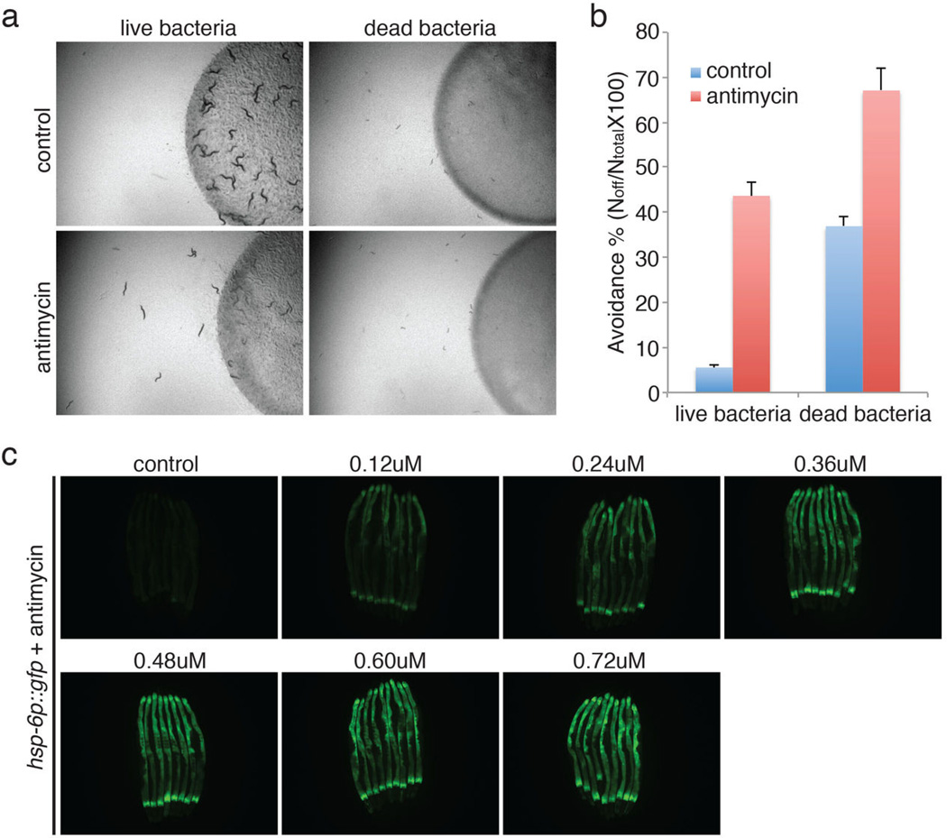
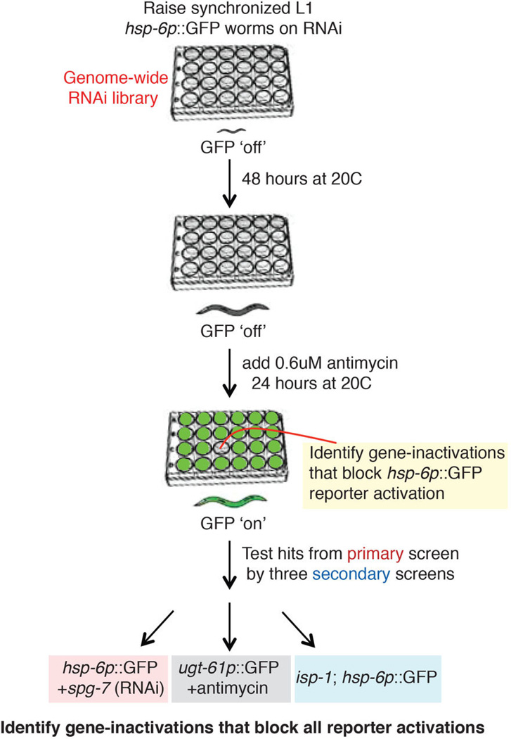
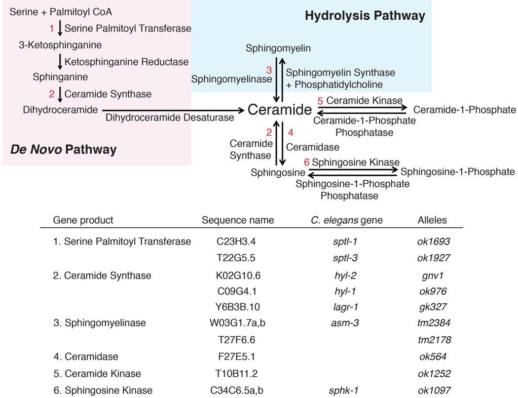
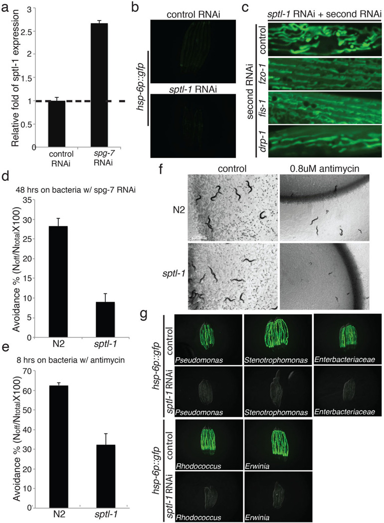
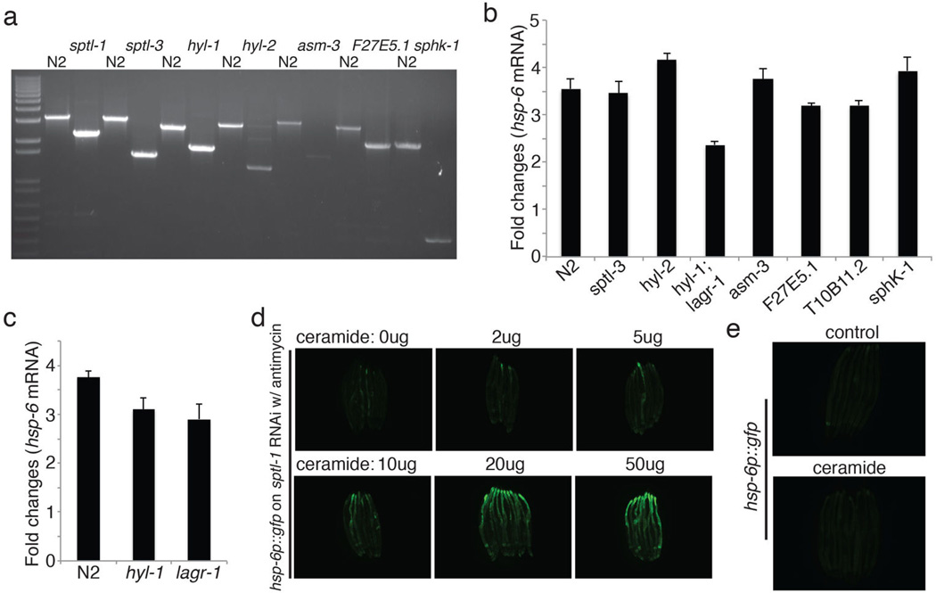
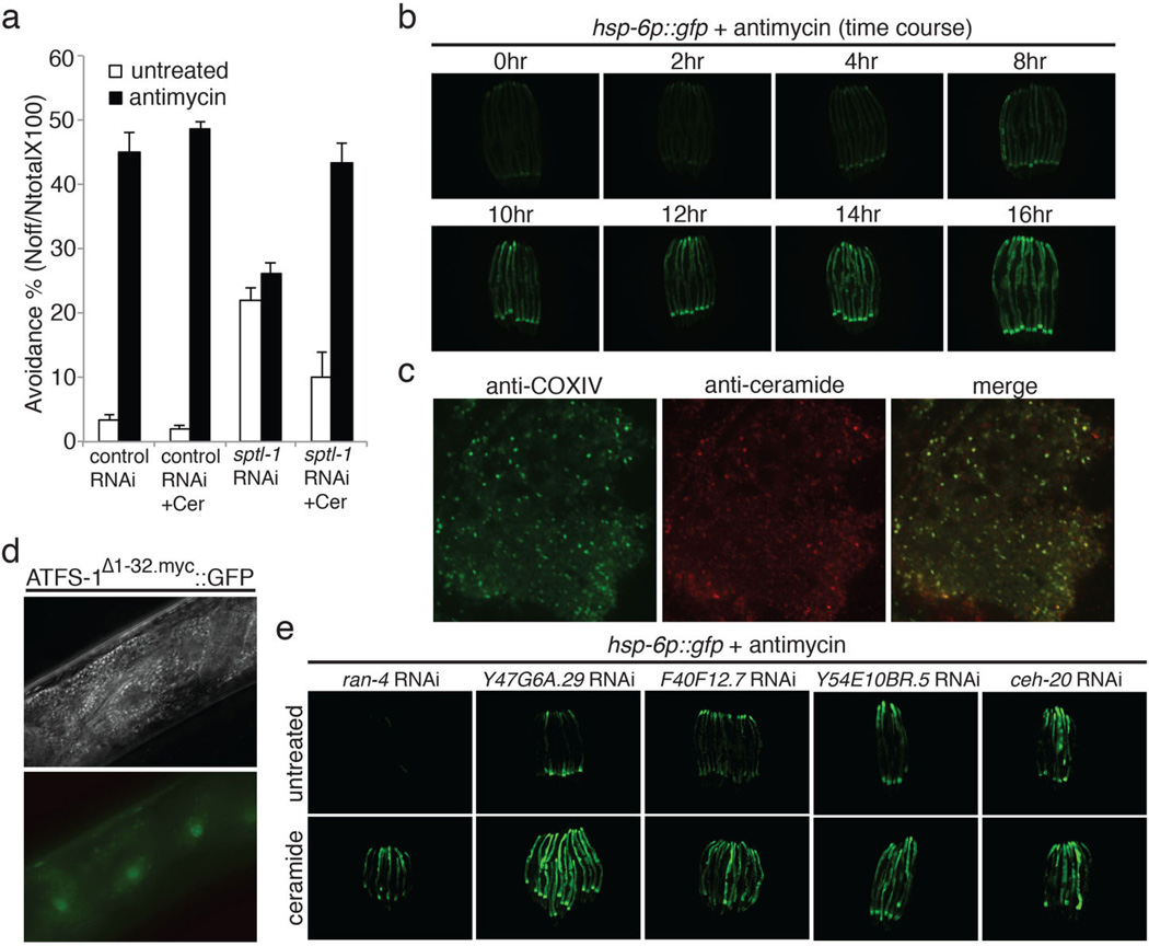
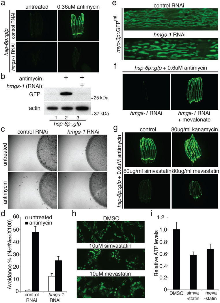
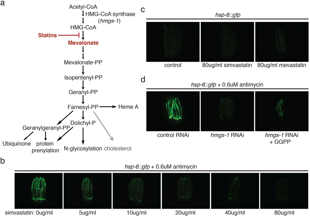
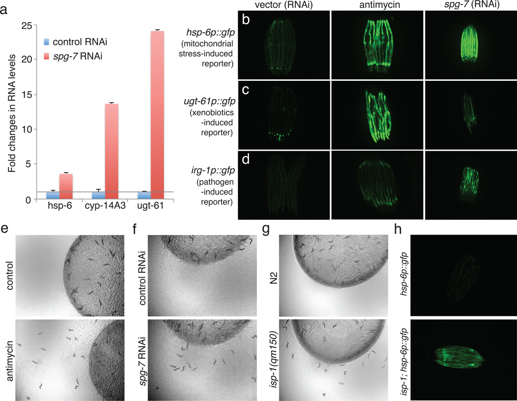
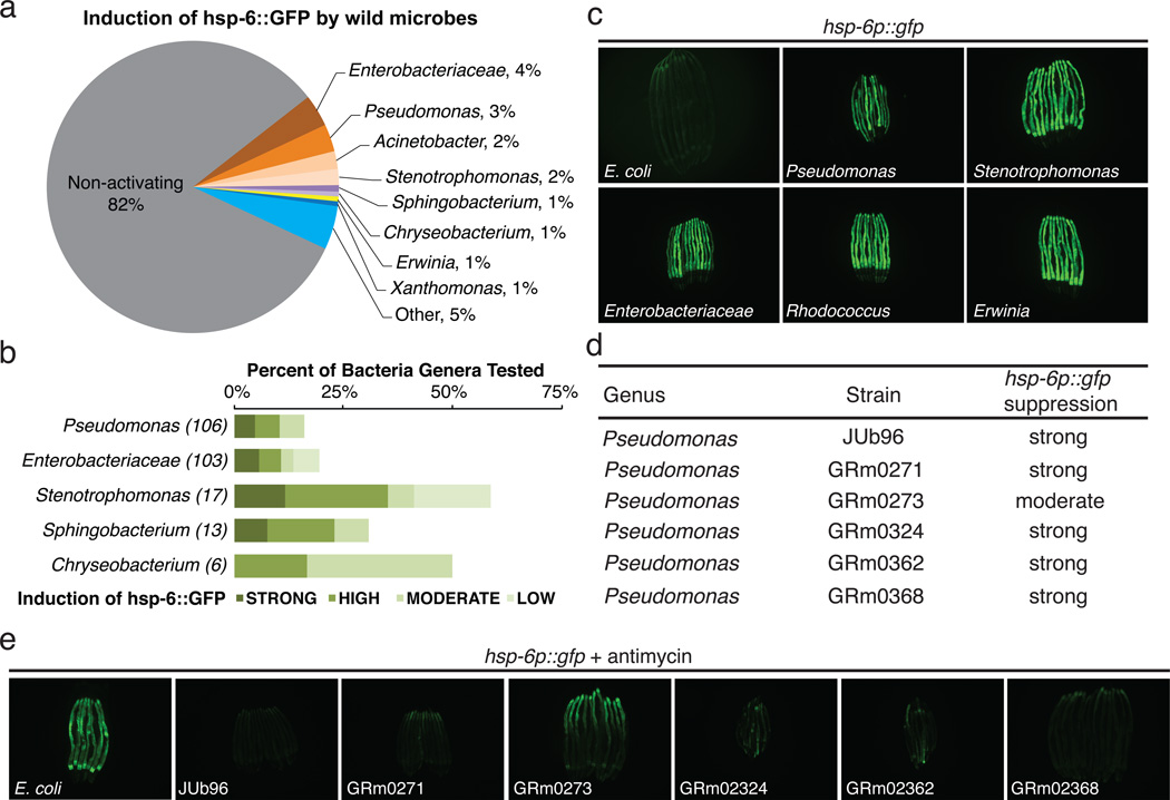
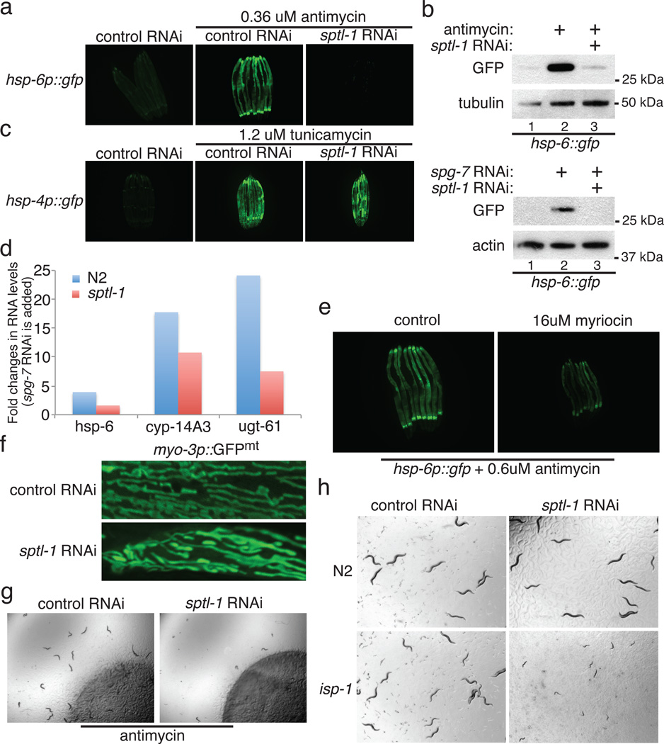
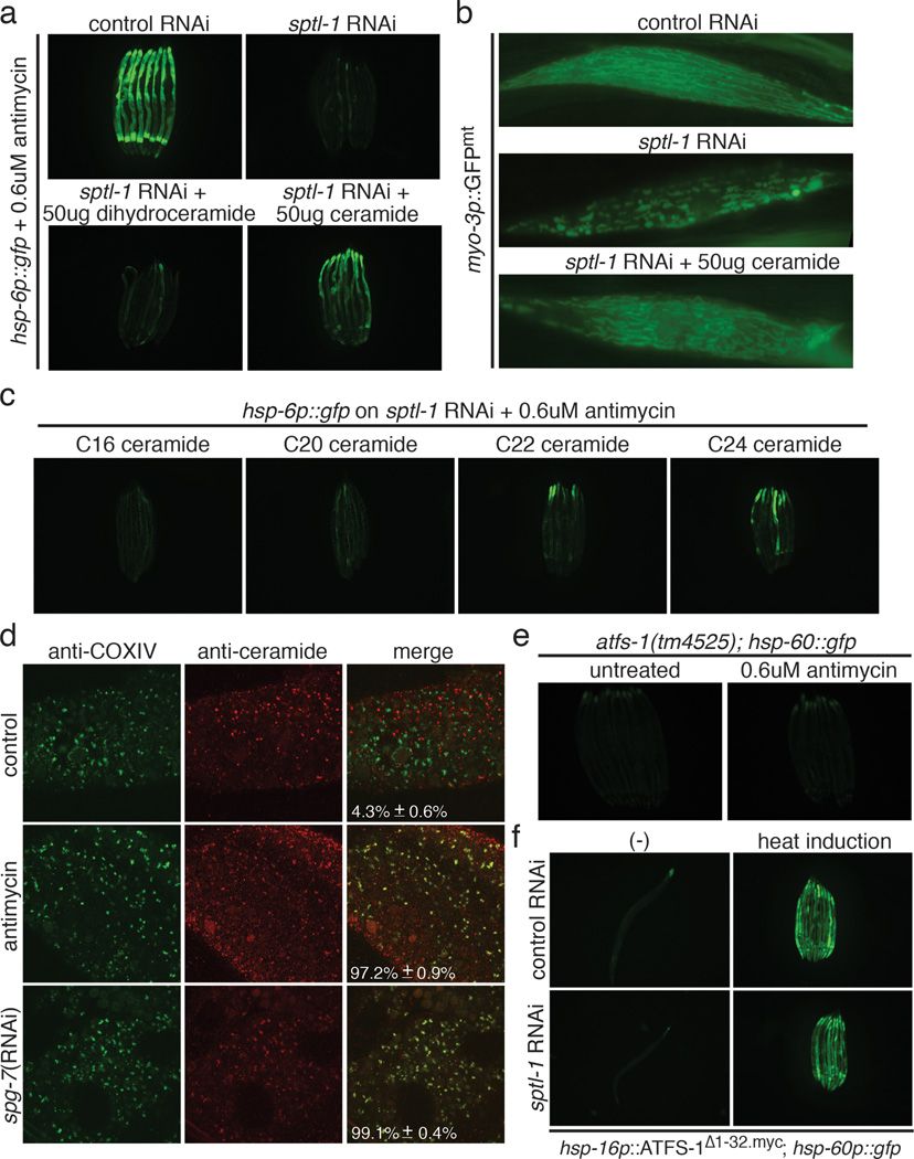
Mitochondrial function is challenged by toxic by-products of metabolism as well as by pathogen attack. Caenorhabditis elegans normally responds to mitochondrial dysfunction with activation of mitochondrial-repair, drug-detoxification and pathogen-response pathways. Here, from a genome-wide RNA interference (RNAi) screen, we identified 45 C. elegans genes that are required to upregulate detoxification, pathogen-response and mitochondrial-repair pathways after inhibition of mitochondrial function by drug-induced or genetic disruption. Animals defective in ceramide biosynthesis are deficient in mitochondrial surveillance, and addition of particular ceramides can rescue the surveillance defects. Ceramide can also rescue the mitochondrial surveillance defects of other gene inactivations, mapping these gene activities upstream of ceramide. Inhibition of the mevalonate pathway, either by RNAi or statin drugs, also disrupts mitochondrial surveillance. Growth of C. elegans with a significant fraction of bacterial species from their natural habitat causes mitochondrial dysfunction. Other bacterial species inhibit C. elegans defence responses to a mitochondrial toxin, revealing bacterial countermeasures to animal defence.


Figures

Extended Data Figure 1
Mitochondrial dysfunction activates homeostatic, detoxification and pathogen responses. a, Drug-induced food avoidance phenotypes on live or dead bacteria. Dead bacteria were obtained by heating bacteria at 90 °C for 30 min. Avoidance behavior was also observed when antimycin was added to dead bacteria, showing that the drug acts directly on C. elegans and is not transformed by the bacteria. b, Quantification of the food avoidance phenotypes of Extended Data Figure 1a (n=4). Error bars represent s.d. c, A dose response of hsp-6p::gfp induction with the addition of antimycin.
Extended Data Figure 2
Diagram of the genome-wide RNAi screen workflow. For the detailed experimental procedure, see Methods.
Extended Data Figure 3
Diagram of the sphingolipid metabolism pathway and the corresponding genes.
Extended Data Figure 4
sptl-1 is required for mitochondrial surveillance. a, A graph showing the fold change in C. elegans sptl-1 transcript level compared to control (n=3). Error bars represent s.d. b, hsp-6p::gfp worms raised on control or sptl-1(RNAi) (n=40). c, Body wall muscle animals expressing a mitochondrially localized GFP reporter. The animals were subjected to sptl-1(RNAi) for 36 hours and transferred onto the second RNAi (control, fzo-1, fis-1 or drp-1). The hyperfusion of mitochondria observed under sptl-1(RNAi) is dependent on the fusion machinery as disruption of mitochondrial fusion by inactivating fzo-1 or fis-1 partially restored the tubular structure. In contrast, inhibition of the gene drp-1 that governs fission led to mitochondrial hyperfusion. The images were taken 36 hours after they were placed on the second RNAi. d, A graph showing the percentage of worms which avoid the spg-7(RNAi) bacteria lawn 48 hours after they were initially placed on the plates (n=4). Error bars represent s.d. e, A graph showing the percentage of worms which avoid the bacteria lawn 8 hours after the addition of antimycin (n=4). Error bars represent s.d. f, wild type N2 or sptl-1(ok1693) mutant animals raised in the presence or absence of 0.8μM antimycin. Photos were taken 4 days after the synchronized L1 worms were placed on the plates. g, hsp-6p::gfp animals were raised on sptl-1(RNAi) for 36 hours and transferred onto a subset of wild microbes. The images were taken after 2 days.
Extended Data Figure 5
Ceramide biosynthesis is required for mitochondrial surveillance. a, Genotyping of the sphingolipid metabolism pathway mutant alleles. b-c, Fold difference in hsp-6 transcript levels in wild type or sphingolipid metabolism pathway mutants (b) and hyl-1 or lagr-1 mutants (c) (n=3). Error bars represent s.d. d, hsp-6p::gfp worms raised on sptl-1(RNAi) in the presence of increasing amounts of ceramide. e, hsp-6p::gfp in the presence or absence of ceramide.
Extended Data Figure 6
Ceramide biogenesis is required for mitochondrial surveillance. a, The percentage of worms that avoid the bacterial lawn 8 hours after the addition of antimycin. Animals were pre-treated with control RNAi, control RNAi with ceramide, sptl-1 RNAi or sptl-1 RNAi with ceramide (n=4). Error bars represent s.d. b, Time course experiment for the induction of hsp-6p::gfp with antimycin. c, Dissected young adults after 4 hours antimycin treatment were stained with anti-COX-IV antibody (red) and anti-ceramide antibody (green). d, Nomarski (upper panel) and fluorescent (lower panel) images of intestinal cells in atfs-1; hsp-16p::atfs-1Δ1-32.myc::gfp transgenic animals. e, hsp-6p::gfp worms raised on indicated RNAi in the presence or absence of ceramide.
Extended Data Figure 7
Inhibition of the mevalonate pathway disrupts mitochondrial surveillance. a, hsp-6p::gfp animals raised on control or hmgs-1(RNAi) in the presence of antimycin. b, Immunoblotting of GFP expressed by hsp-6p::gfp animals, with or without antimycin. c, Antimycin induced food avoidance in control or hmgs-1(RNAi) animals. d, Quantification of food avoidance (n=4). Error bars represent s.d. e, Body wall muscle of control or hmgs-1(RNAi) animals expressing a mitochondrially localized GFP reporter. f, hsp-6p::gfp animals raised on hmgs-1(RNAi), or hmgs-1(RNAi) with addition of mevalonate exposed to antimycin. g, hsp-6p::gfp animals treated with antimycin after pre-treatment with simvastatin or mevastatin. h, Mitochondrial immunostaining in HEK293T cells. i, ATP levels in C2C12 myotubes after treating with simvastatin or mevastatin (n=3). Error bars represent s.d.
Extended Data Figure 8
a, Diagram of the mevalonate pathway for the biosynthesis of cholesterol, ubiquinone and heme A, and protein N-glycosylation and prenylation. b, hsp-6p::gfp animals treated with antimycin after pre-treatment with increasing concentration of simvastatin. c, hsp-6p::gfp animals with mock, 80ug/ml simvastatin or 80ug/ml mevastatin treatment. d, hsp-6p::gfp animals raised on control RNAi, hmgs-1 RNAi, or hmgs-1 RNAi with the addition of geranylgeranyl pyrophoshate. The animals were then treated with antimycin to induce mitochondrial damage. Statin toxicity has been proposed to be caused by the inhibition of Rab prenylation. Geranylgeranyl pyrophosphate (GGPP), a precursor of protein prenylation rescued the statin side effect in cell culture. GGPP also partially rescued the deficiency of mitochondrial surveillance and activated hsp-6p::gfp in antimycin-treated hmgs-1(RNAi) animals.
Extended Data Figure 9
Mitochondrial immunostaining in HEK293T cells. The cells were treated with DMSO, 10uM simvastatin or 10uM mevastatin for 2 days.
Figure 1
Figure 1
Mitochondrial dysfunction activates homeostatic, detoxification and pathogen responses. a, mitochondrial chaperone and detoxification transcripts in control vs. spg-7(RNAi) (n=3). Error bars represent s.d. b-d, hsp-6p::gfp (b), ugt-61p::gfp (c), and irg-1p::gfp (d) animals raised on control E. coli, or E. coli with antimycin or spg-7 (RNAi). e-g, Drug-, RNAi- or mutant allele-based mitochondrial inhibition causes food avoidance. h, wild type hsp-6p::gfp and isp-1(qm150); hsp-6p::gfp animals.
Figure 2
Figure 2
Some bacteria from the C. elegans natural habitat antagonize the mitochondria. a, Genera that induce hsp-6p::gfp [100 of 560 tested microbes (18%)]. b, Proportion of bacterial genera tested that cause mitochondrial dysfunction and hsp-6p::gfp induction. c, hsp-6p::gfp animals raised on E. coli or natural microbial species. d, Six bacterial strains that render C. elegans defective in hsp-6 response to antimycin. e, hsp-6p::gfp animals exposed to antimycin and raised on control E. coli or the six microbial strains.
Figure 3
Figure 3
The serine palmitoyl transferase sptl-1 is required for mitochondrial surveillance. a, hsp-6p::gfp animals raised on control or sptl-1(RNAi) in the presence or absence of antimycin. b, GFP immunoblot expressed by hsp-6p::gfp animals in the presence or absence of antimycin or spg-7(RNAi). c, hsp-4p::gfp animals raised on control or sptl-1(RNAi) in the presence or absence of the ER drug tunicamycin. d, C. elegans hsp-6 and xenobiotic detoxification gene transcripts in control or sptl-1(ok1693) mutant animals after exposure to spg-7(RNAi) (n=2). e, hsp-6p::gfp animals raised in the presence or absence of myriocin. f, Body wall muscle of sptl-1(RNAi) compared to wild type animals expressing a mitochondrially localized GFP reporter. g, Food avoidance phenotypes for control or sptl-1(RNAi) animals treated with antimycin. h, Wild type or isp-1(qm150) raised on control or sptl-1(RNAi).
Figure 4
Figure 4
Ceramide biogenesis is required for mitochondrial surveillance. a, hsp-6p::gfp animals exposed to antimycin in the presence or absence of dihydroceramide or ceramide. b, Body wall muscle of animals expressing a mitochondrially localized GFP reporter in sptl-1(RNAi) with and without added ceramide. c, hsp-6p::gfp sptl-1(RNAi) plus antimycin with different ceramide species. d, Dissected animals after antimycin or spg-7(RNAi) treatment were stained with anti-COX-IV (red) and anti-ceramide antibodies (green). Quantification represents proportion of mitochondria with contact of mitochondrial and ceramide staining (mean ± s.d., n=3). e, atfs-1(tm4525); hsp-60p::gfp animals alone or with antimycin. f, atfs-1(tm4525); hsp-16p::ATFS-1Δ1-32.myc; hsp-60p::gfp animals raised on control or sptl-1(RNAi).

Comment in

References

    1. Melo JA, Ruvkun G. Inactivation of conserved C. elegans genes engages pathogen- and xenobiotic-associated defenses. Cell. 2012;149:452–466. - PMC - PubMed
    1. Runkel ED, Liu S, Baumeister R, Schulze E. Surveillance-activated defenses block the ROS-induced mitochondrial unfolded protein response. PLoS genetics. 2013;9:e1003346. - PMC - PubMed
    1. Benedetti C, Haynes CM, Yang Y, Harding HP, Ron D. Ubiquitin-like protein 5 positively regulates chaperone gene expression in the mitochondrial unfolded protein response. Genetics. 2006;174:229–239. - PMC - PubMed
    1. Haynes CM, Petrova K, Benedetti C, Yang Y, Ron D. ClpP mediates activation of a mitochondrial unfolded protein response in C. elegans. Developmental cell. 2007;13:467–480. - PubMed
    1. Haynes CM, Yang Y, Blais SP, Neubert TA, Ron D. The matrix peptide exporter HAF-1 signals a mitochondrial UPR by activating the transcription factor ZC376.7 in C. elegans. Molecular cell. 2010;37:529–540. - PMC - PubMed

Publication types

MeSH terms

Substances