. 2014 Apr 2;9(4):e93373.
doi: 10.1371/journal.pone.0093373. eCollection 2014.

Epidermal or dermal specific knockout of PHD-2 enhances wound healing and minimizes ischemic injury

Affiliations

Epidermal or dermal specific knockout of PHD-2 enhances wound healing and minimizes ischemic injury

Andrew S Zimmermann et al. PLoS One. .

Abstract

Introduction: Hypoxia-inducible factor (HIF)-1α, part of the heterodimeric transcription factor that mediates the cellular response to hypoxia, is critical for the expression of multiple angiogenic growth factors, cell motility, and the recruitment of endothelial progenitor cells. Inhibition of the oxygen-dependent negative regulator of HIF-1α, prolyl hydroxylase domain-2 (PHD-2), leads to increased HIF-1α and mimics various cellular and physiological responses to hypoxia. The roles of PHD-2 in the epidermis and dermis have not been clearly defined in wound healing.

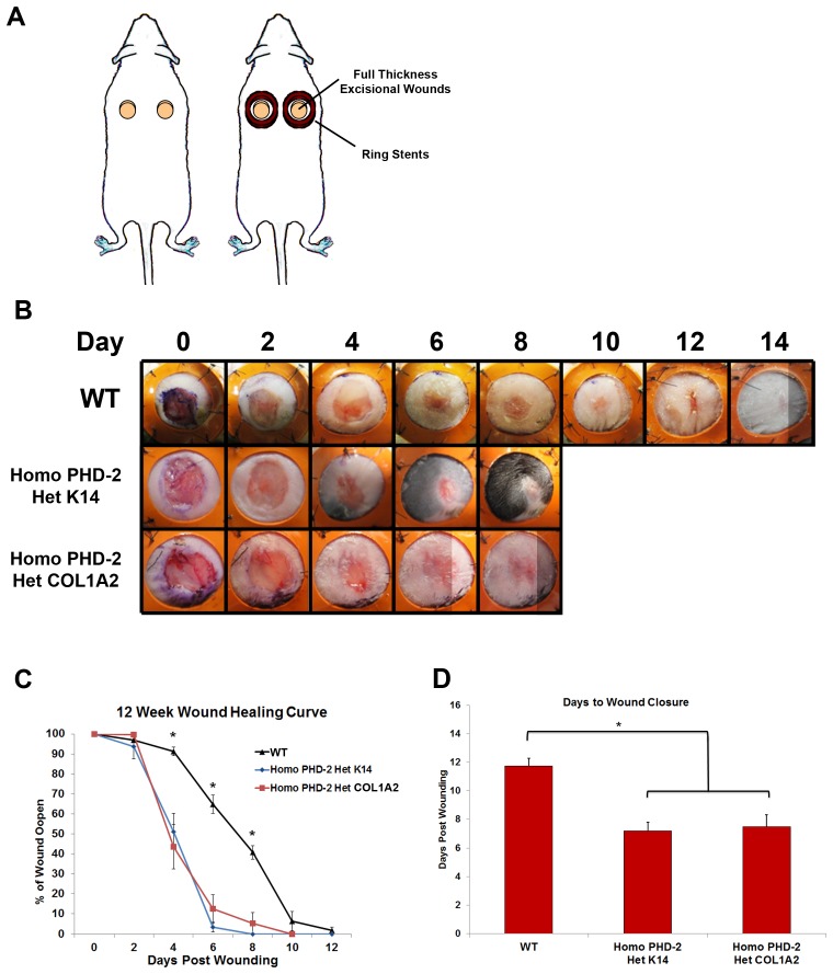
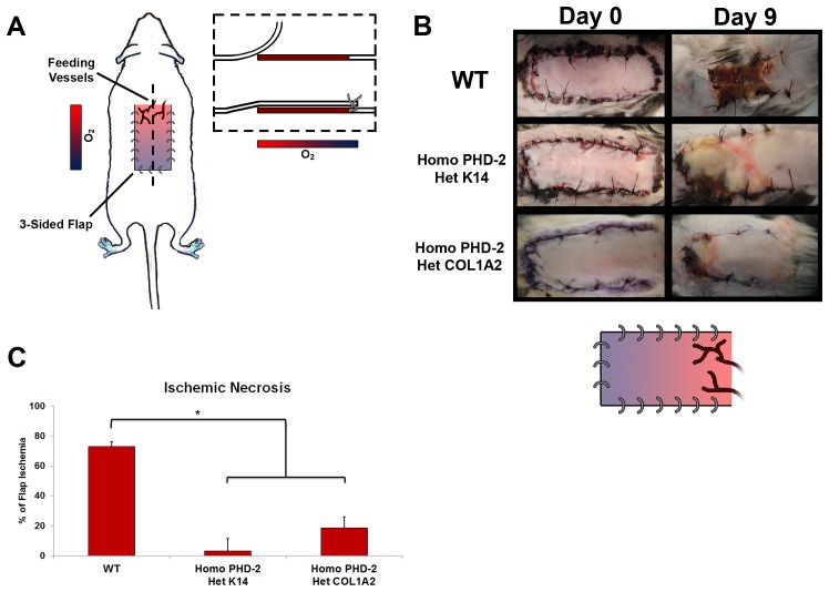
Methods: Epidermal and dermal specific PHD-2 knockout (KO) mice were developed in a C57BL/6J (wild type) background by crossing homozygous floxed PHD-2 mice with heterozygous K14-Cre mice and heterozygous Col1A2-Cre-ER mice to get homozygous floxed PHD-2/heterozygous K14-Cre and homozygous floxed PHD-2/heterozygous floxed Col1A2-Cre-ER mice, respectively. Ten to twelve-week-old PHD-2 KO and wild type (WT) mice were subjected to wounding and ischemic pedicle flap model. The amount of healing was grossly quantified with ImageJ software. Western blot and qRT-PCR was run on protein and RNA from primary cells cultured in vitro.

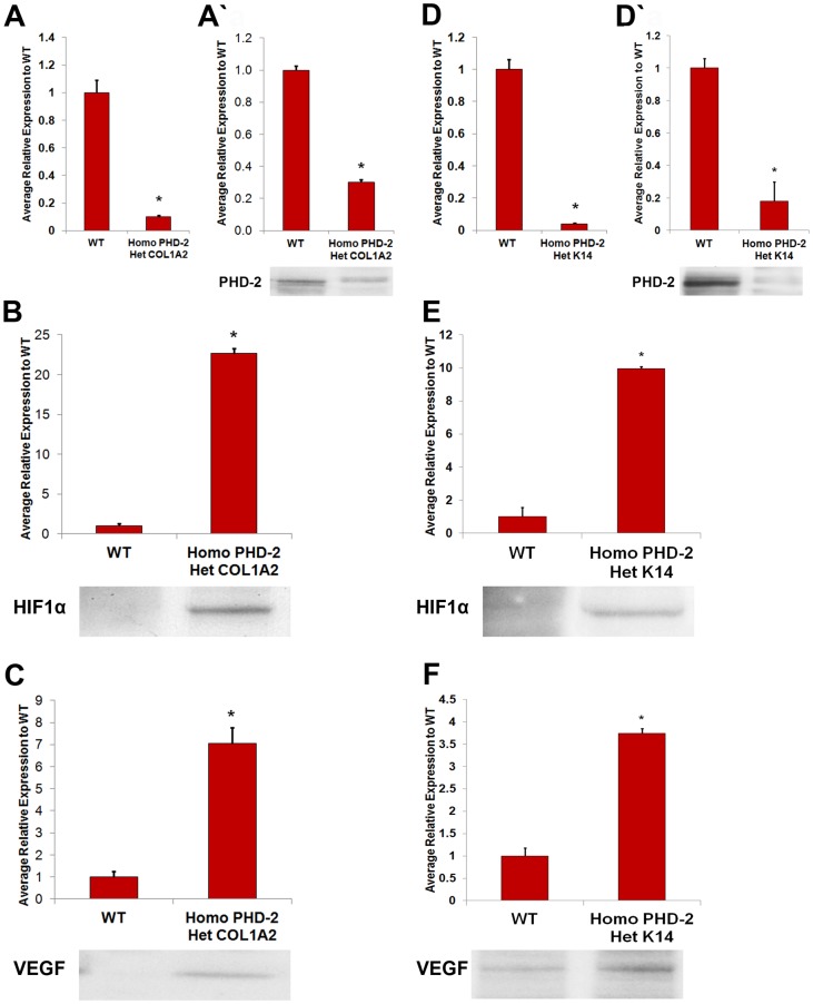
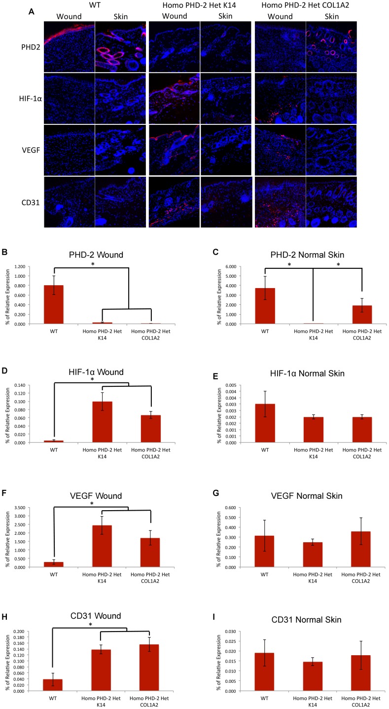
Results: qRT-PCR demonstrated a significant decrease of PHD-2 in keratinocytes and fibroblasts derived from tissue specific KO mice relative to control mice (*p<0.05). Western blot analysis showed a significant increase in HIF-1α and VEGF protein levels in PHD-2 KO mice relative to control mice (*p<0.05). PHD-2 KO mice showed significantly accelerated wound closure relative to WT (*p<0.05). When ischemia was analyzed at day nine post-surgery in a flap model, the PHD-2 tissue specific knockout mice showed significantly more viable flaps than WT (*p<0.05).

Conclusions: PHD-2 plays a significant role in the rates of wound healing and response to ischemic insult in mice. Further exploration shows PHD-2 KO increases cellular levels of HIF-1α and this increase leads to the transcription of downstream angiogenic factors such as VEGF.


Conflict of interest statement

Competing Interests: The authors have declared that no competing interests exist.

Figures

Figure 1
Figure 1. In vitro analysis of PHD-2 knockout and protein quantification.
A) Quantitative polymerase chain reaction for PHD-2 knockout in heterozygous Col1α2-Cre-ER/homozygous floxed PHD-2 fibroblasts compared to wild type fibroblasts (*p<0.05). A’) Western blot data for PHD-2 knockout in fibroblasts of heterozygous Col1α2-Cre-ER/homozygous floxed PHD-2 mice compared to wild type mice (*p<0.05). B) Western blot data for HIF-1α in fibroblasts of heterozygous Col1α2-Cre-ER/homozygous floxed PHD-2 mice compared to wild type mice (*p<0.05). C) Western blot data for VEGF in fibroblasts of heterozygous Col1α2-Cre-ER/homozygous floxed PHD-2 mice compared to wild type mice (*p<0.05). D) Quantitative polymerase chain reaction for PHD-2 knockout in heterozygous K14-Cre/homozygous floxed PHD-2 keratinocytes compared to wild type keratinocytes (*p<0.05). D’) Western blot data for PHD-2 knockout in keratinocytes of heterozygous K14-Cre/homozygous floxed PHD-2 mice compared to wild type mice (*p<0.05). E) Western blot data for HIF-1α in keratinocytes of heterozygous K14-Cre/homozygous floxed PHD-2 mice compared to wild type mice (*p<0.05). F) Western blot data for VEGF in keratinocytes of heterozygous K14-Cre/homozygous floxed PHD-2 mice compared to wild type mice (*p<0.05).
Figure 2
Figure 2. In vivo data from humanized wound healing model.
A) Schematic of stented full-thickness excisional wound model. B) Representative pictures of wounds from days 0–14 for wild type, heterozygous K14-Cre/homozygous floxed PHD-2, and heterozygous Col1α2-Cre-ER/homozygous floxed PHD-2 mice. C) Twelve week wound healing curve showing the wound closure rate for wild type, heterozygous K14-Cre/homozygous floxed PHD-2, and heterozygous Col1α2-Cre-ER/homozygous floxed PHD-2 mice with significant differences on days 4, 6, and 8 (*p<0.05). D) Graph representing days to wound closure with a significant difference in days to closure between the wild type and the heterozygous K14-Cre/homozygous floxed PHD-2 and heterozygous Col1α2-Cre-ER/homozygous floxed PHD-2 mice (*p<0.05).
Figure 3
Figure 3. In vivo data from ischemic pedicle flap model.
A) Schematic of ischemic pedicle flap model. B) Representative figures taken at day 0 and 9 of ischemic flaps for wild type, heterozygous K14-Cre/homozygous floxed PHD-2, and heterozygous Col1α2-Cre-ER/homozygous floxed PHD-2 mice. A schematic of tissue reperfusion gradient is also shown. C) Graphical representation of total flap ischemia with a significant difference (*p<0.05) between the wild type and the heterozygous K14-Cre/homozygous floxed PHD-2, and heterozygous Col1α2-Cre-ER/homozygous floxed PHD-2 mice.
Figure 4
Figure 4. Immunofluorescence of wounded tissue and normal unwounded skin.
A) Representative images for immunofluorescent staining of PHD-2, HIF-1α, VEGF, and CD31 on healed wounded skin and adjacent normal unwounded skin of wild type, heterozygous K14-Cre/homozygous floxed PHD-2, and heterozygous Col1α2-Cre-ER/homozygous floxed PHD-2 mice at 400× magnification. B) Relative protein immunolocalization of PHD-2 in healed wounds of wild type, heterozygous K14-Cre/homozygous floxed PHD-2, and heterozygous Col1α2-Cre-ER/homozygous floxed PHD-2 mice (*p<0.05). C) Relative protein immunolocalization of PHD-2 in adjacent normal unwounded skin of wild type, heterozygous K14-Cre/homozygous floxed PHD-2, and heterozygous Col1α2-Cre-ER/homozygous floxed PHD-2 mice (*p<0.05). D) Relative protein immunolocalization of HIF-1α in healed wounds of wild type, heterozygous K14-Cre/homozygous floxed PHD-2, and heterozygous Col1α2-Cre-ER/homozygous floxed PHD-2 mice (*p<0.05). E) Relative protein immunolocalization of HIF-1α in adjacent normal unwounded skin of wild type, heterozygous K14-Cre/homozygous floxed PHD-2, and heterozygous Col1α2-Cre-ER/homozygous floxed PHD-2 mice. F) Relative protein immunolocalization of VEGF in healed wounds of wild type, heterozygous K14-Cre/homozygous floxed PHD-2, and heterozygous Col1α2-Cre-ER/homozygous floxed PHD-2 mice (*p<0.05). G) Relative protein immunolocalization of VEGF in adjacent normal unwounded skin of wild type, heterozygous K14-Cre/homozygous floxed PHD-2, and heterozygous Col1α2-Cre-ER/homozygous floxed PHD-2 mice. H) Relative immunolocalization of CD31 in healed wounds of wild type, heterozygous K14-Cre/homozygous floxed PHD-2, and heterozygous Col1α2-Cre-ER/homozygous floxed PHD-2 mice (*p<0.05). I) Relative immunolocalization of CD31 in adjacent normal unwounded skin of wild type, heterozygous K14-Cre/homozygous floxed PHD-2, and heterozygous Col1α2-Cre-ER/homozygous floxed PHD-2 mice.

References

    1. Sen CK, Gordillo GM, Roy S, Kirsner R, Lambert L, et al. (2009) Human skin wounds: a major and snowballing threat to public health and the economy. Wound Repair Regen 17: 763–771. - PMC - PubMed
    1. Kranke P, Bennett MH, Martyn-St James M, Schnabel A, Debus SE (2012) Hyperbaric oxygen therapy for chronic wounds. Cochrane Database Syst Rev 4: CD004123. - PubMed
    1. Fonder MA, Lazarus GS, Cowan DA, Aronson-Cook B, Kohli AR, et al. (2008) Treating the chronic wound: A practical approach to the care of nonhealing wounds and wound care dressings. J Am Acad Dermatol 58: 185–206. - PubMed
    1. Kirsner RS, Warriner R, Michela M, Stasik L, Freeman K (2010) Advanced biological therapies for diabetic foot ulcers. Arch Dermatol 146: 857–862. - PubMed
    1. Driver VR, Fabbi M, Lavery LA, Gibbons G (2010) The costs of diabetic foot: the economic case for the limb salvage team. J Vasc Surg 52: 17S–22S. - PubMed

Substances