. 2014 Apr 2;9(4):e93228.
doi: 10.1371/journal.pone.0093228. eCollection 2014.

Solution structure of CXCL5--a novel chemokine and adipokine implicated in inflammation and obesity

Affiliations

Solution structure of CXCL5--a novel chemokine and adipokine implicated in inflammation and obesity

Krishna Mohan Sepuru et al. PLoS One. .

Abstract

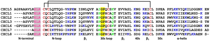
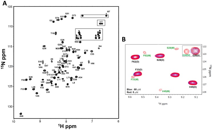
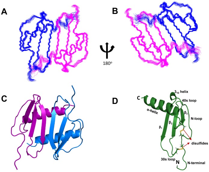
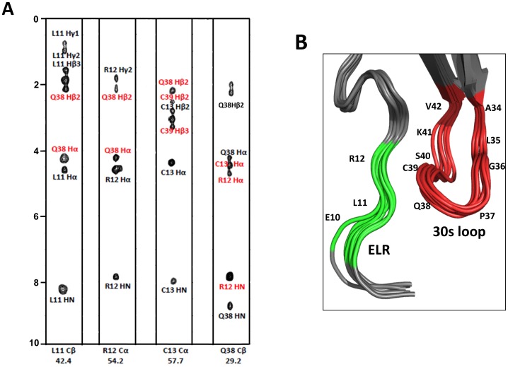
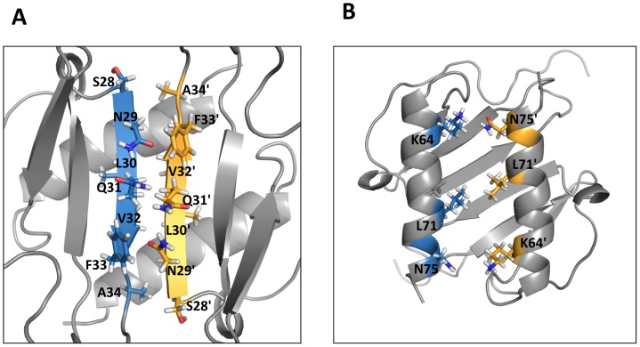
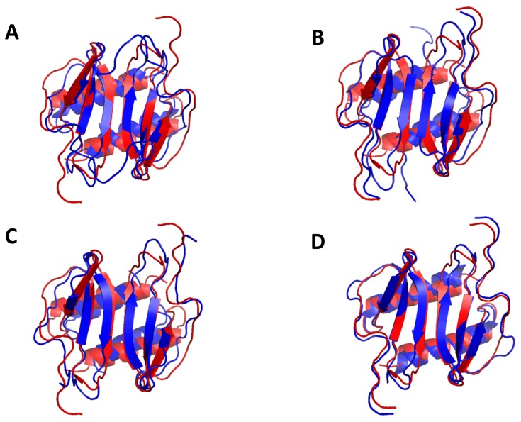
The chemokine CXCL5 is selectively expressed in highly specialized cells such as epithelial type II cells in the lung and white adipose tissue macrophages in muscle, where it mediates diverse functions from combating microbial infections by regulating neutrophil trafficking to promoting obesity by inhibiting insulin signaling. Currently very little is known regarding the structural basis of how CXCL5 mediates its novel functions. Towards this missing knowledge, we have solved the solution structure of the CXCL5 dimer by NMR spectroscopy. CXCL5 is a member of a subset of seven CXCR2-activating chemokines (CAC) that are characterized by the highly conserved ELR motif in the N-terminal tail. The structure shows that CXCL5 adopts the typical chemokine fold, but also reveals several distinct differences in the 30 s loop and N-terminal residues; not surprisingly, crosstalk between N-terminal and 30 s loop residues have been implicated as a major determinant of receptor activity. CAC function also involves binding to highly sulfated glycosaminoglycans (GAG), and the CXCL5 structure reveals a distinct distribution of positively charged residues, suggesting that differences in GAG interactions also influence function. The availability of the structure should now facilitate the design of experiments to better understand the molecular basis of various CXCL5 functions, and also serve as a template for the design of inhibitors for use in a clinical setting.


Conflict of interest statement

Competing Interests: The authors have declared that no competing interests exist.

Figures

Figure 1
Figure 1. Sequence alignment of CXCR2-activating chemokines.
The conserved ELR, cysteine, and GAG-binding residues are highlighted in magenta, red, and blue, respectively. In the 30 s loop region, GP motif and residues that are aligned with I35 and K41 are highlighted in yellow and green, respectively.
Figure 2
Figure 2. Structural Characterization of the CXCL5 dimer.
(A) 15N-1H HSQC 800 MHz NMR spectrum of the CXCL5 dimer at pH 6.0 and 25°C. The spectrum shows excellent chemical shift dispersion indicating a well-folded single species with no evidence of heterogeneity. The NH2 resonances of asparagines and glutamines are boxed. (B) Sections of the 15N-1H HSQC spectra at pH 7.5 and 25°C at 5 μM (red) and 80 μM (blue). A new set of peaks corresponding to the monomer is evident in the 5 μM spectrum. The equilibrium constant was calculated to be ∼0.3 μM on the basis of monomer and dimer intensities.
Figure 3
Figure 3. NMR Solution Structure of CXCL5 dimer.
Panels A and B show the superposition of the backbone atoms N, Cα and CO of residues 10 to 78 for the calculated twenty structures in two orientations. The polypeptide backbone is colored in blue and magenta for the two monomers. (C) Ribbon representation of the CXCL5 [10–78] structure. The protein dimer comprises of a six-stranded antiparallel β-sheet and a pair of α-helices. (D) Ribbon representation of CXCL5 monomer. The monomer consists of three antiparallel beta strands and an alpha helix; the disulfide bonds are shown in yellow.
Figure 4
Figure 4. Interactions between 30 s loop and ELR residues.
(A) Strip plots of the 13C-edited NOESY spectra from L11Cβ, R12Cα, C13Cα, and Q38Cβ. The intra and inter-residue NOEs are labeled in black and red, respectively. (B) Zoomed-in view of the ELR motif (in green) and 30 s loop (in red) residues.
Figure 5
Figure 5. A schematic of dimer-interface interactions.
(A) β1/β1′ and (B) α1/α1′ residues that stabilize the dimer interface are highlighted. The interface residues in the monomer units are represented in blue and yellow, respectively.
Figure 6
Figure 6. Structural comparison between CXCL5 and other CXCR2-activating chemokines.
(A) Superposition of CXCL5 [8–78] (red) on CXCL1, CXCL2, CXCL7, and CXCL8 (blue) in two different orientations. The superposition was optimized using residues 8 to 78 of CXCL5, residues 8 to 73 of CXCL1, residues 6 to 68 of CXCL2, residues 7 to 68 of CXCL7, and residues 8 to 72 of CXCL8.
Figure 7
Figure 7. Stability features from amide exchange.
HSQC spectra of 100 μM CXCL5 in 50 mM sodium phosphate pH 6.0 after initiating exchange with D2O after ∼8 min and after ∼24 hrs. The panel C shows the plot of ΔGHX calculated from the amide exchange data.
Figure 8
Figure 8. Backbone dynamics.
A plot of the backbone {1H}-15N NOE values as a function of residue number. The data show that the terminal residues are flexible and also the N-loop and the turn residues are more dynamic compared to the helical and strand residues.
Figure 9
Figure 9. Electrostatic representation of CXCR2-activating chemokines.
The upper panels show the helical surface that highlight distribution of the GAG-binding residues and the lower panels show the opposite β-sheet surface after a 180° flip.

References

    1. Zlotnik A, Yoshie O (2000) Chemokines: a new classification system and their role in immunity. Immunity 12: 121–127. - PubMed
    1. Fernandez EJ, Lolis E (2002) Structure, function, and inhibition of chemokines. Annu Rev Pharmacol Toxicol 42: 469–499. - PubMed
    1. Bonecchi R, Galliera E, Borroni EM, Corsi MM, Locati M, et al. (2009) Chemokines and chemokine receptors: an overview. Front Biosci 14: 540–551. - PubMed
    1. O'Hayre M, Salanga CL, Handel TM, Allen SJ (2008) Chemokines and cancer: migration, intracellular signalling and intercellular communication in the microenvironment. Biochem J 409: 635–649. - PubMed
    1. Stillie R, Farooq SM, Gordon JR, Stadnyk AW (2009) The functional significance behind expressing two IL-8 receptor types on PMN. J Leukoc Biol 86: 529–543. - PubMed

Publication types