. 2014 Apr 2;34(14):4801-8.
doi: 10.1523/JNEUROSCI.4393-13.2014.

14-3-3 proteins are required for hippocampal long-term potentiation and associative learning and memory

Affiliations

14-3-3 proteins are required for hippocampal long-term potentiation and associative learning and memory

Haifa Qiao et al. J Neurosci. .

Abstract

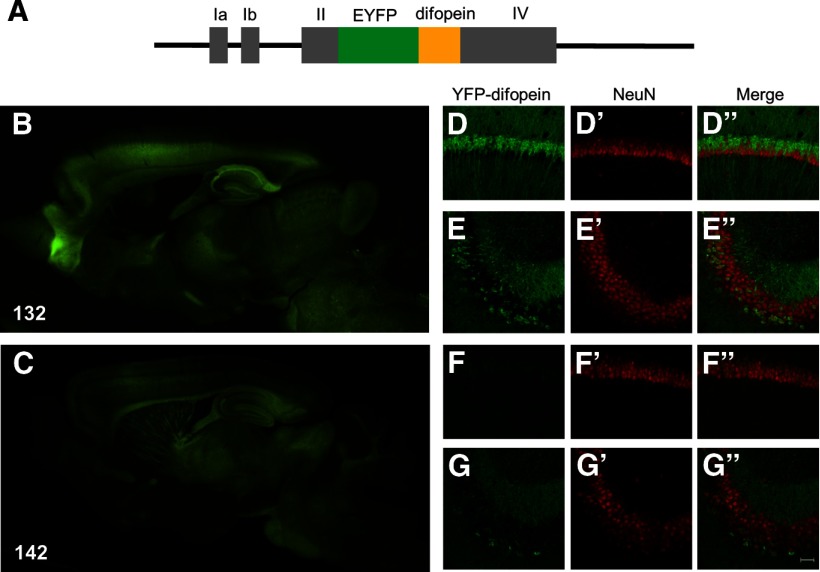
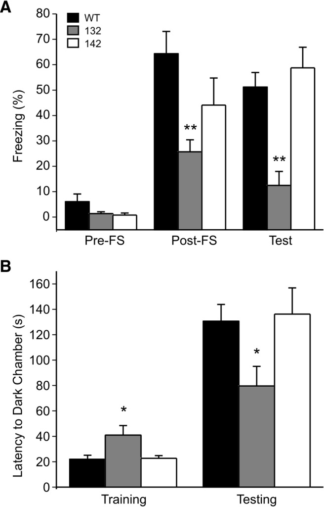
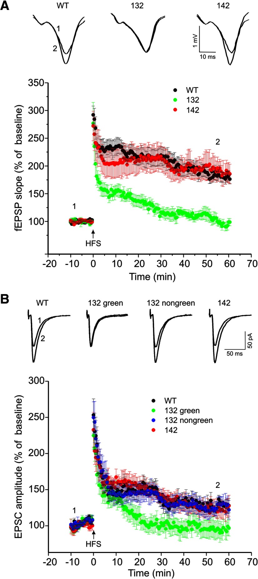
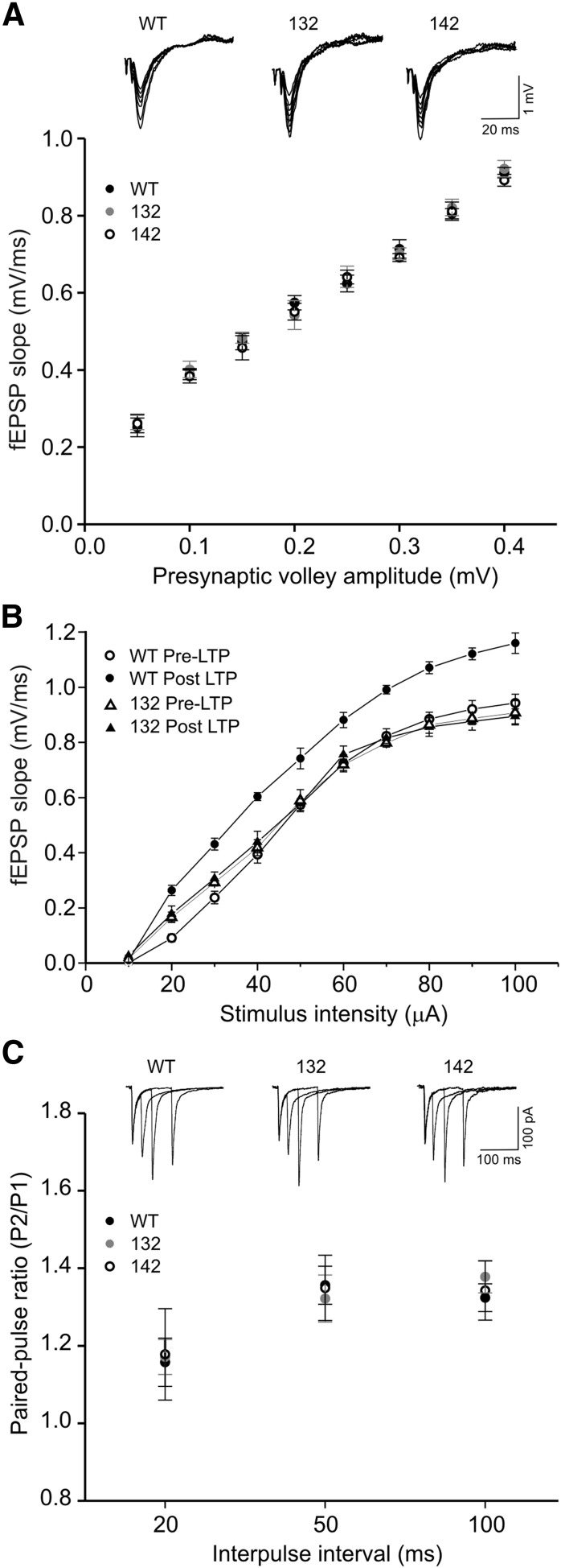
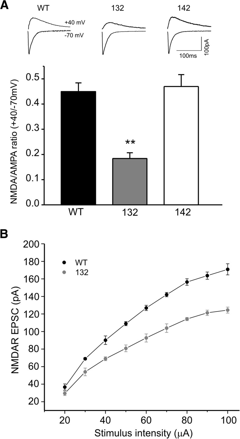
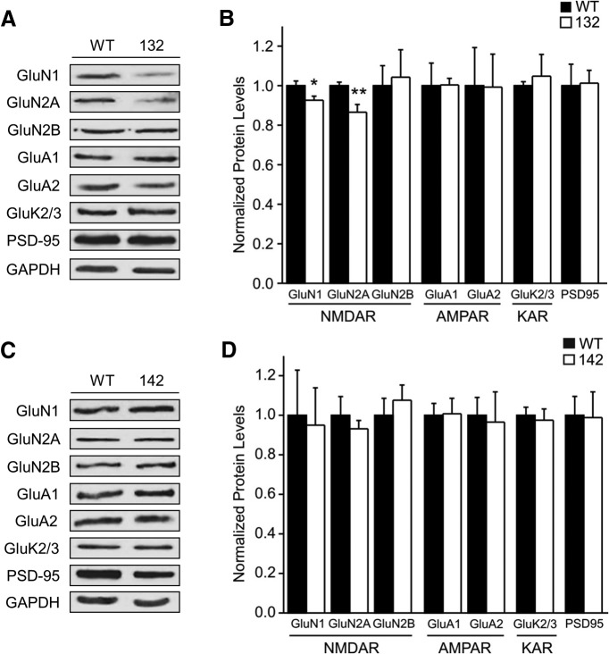
14-3-3 is a family of regulatory proteins highly expressed in the brain. Previous invertebrate studies have demonstrated the importance of 14-3-3 in the regulation of synaptic functions and learning and memory. However, the in vivo role of 14-3-3 in these processes has not been determined using mammalian animal models. Here, we report the behavioral and electrophysiological characterization of a new animal model of 14-3-3 proteins. These transgenic mice, considered to be a 14-3-3 functional knock-out, express a known 14-3-3 inhibitor in various brain regions of different founder lines. We identify a founder-specific impairment in hippocampal-dependent learning and memory tasks, as well as a correlated suppression in long-term synaptic plasticity of the hippocampal synapses. Moreover, hippocampal synaptic NMDA receptor levels are selectively reduced in the transgenic founder line that exhibits both behavioral and synaptic plasticity deficits. Collectively, our findings provide evidence that 14-3-3 is a positive regulator of associative learning and memory at both the behavioral and cellular level.

Keywords: 14-3-3 proteins; NMDA receptors; fear conditioning; long-term potentiation; passive avoidance.


Figures

Figure 1.
Figure 1.
Transgene expressions in the brains of two different founder lines of 14-3-3 FKO mice. A, Schematic representation of the Thy1-YFP-difopein expression cassette used in this study. The gray bars represent the untranslated exons of the Thy-1.2 gene. B, C, Detection of YFP-difopein fusion proteins by fluorescence microscopy in sagittal brain sections of 132 and 142 founder line mice. D–G, Confocal images showing YFP-difopein signals in the hippocampal CA1 (132, D; 142, F) and CA3 (132, E; 142, G) subregions of 132 and 142 founder line mice. Brain sections were immunostained with a NeuN antibody to visualize neuronal nuclei (D′, E′, F′, G′). Transgene expression in NeuN positive neurons is shown by merged images (132, D” and E”; 142, F” and G”). Scale bar, 40 μm.
Figure 2.
Figure 2.
Founder-specific deficits of learning and memory behaviors in 14-3-3 FKO mice. Hippocampal-dependent associative learning and memory were assessed using contextual fear conditioning (A) and passive avoidance testing (B). A, Compared with WT, the 132 founder line mice showed significantly less freezing immediately post-FS and 24 h later when re-exposed to the same context (Test). 142 founder line mice showed no difference from the WT in the freezing responses. (WT, n = 7; 132, n = 10; 142, n = 5). B, Compared with WT, the 132 mice had a shorter latency to enter the dark chamber when tested 24 h (Testing) after footshock (Training). There was no difference between 142 mice and the WT in their latency to the dark side (WT, n = 21; 132, n = 18; 142, n = 10). All data are represented as mean ± SEM, with *p < 0.05, **p < 0.01, one-way ANOVA.
Figure 3.
Figure 3.
Founder-specific impairment of hippocampal LTP in 14-3-3 FKO mice. A, HFS-induced LTP at Sch-CA1 synapses was measured by field recording in WT, 132, or 142 founder line mice. Representative fEPSP traces before (at position 1) and after (at position 2) the induction of LTP are shown on the top (WT, n = 15; 132, n = 12; 142, n = 10). B, Hippocampal LTP was assessed by whole-cell recording in CA1 pyramidal cells from WT and the two founder lines of 14-3-3 FKO mice. For 132 founder line mice, LTP of EPSCs was evaluated separately in neurons with (132 green) and without (132 non-green) fluorescence. Shown on the top are representative EPSC traces recorded from CA1 pyramidal neurons before and after LTP induction. (WT, n = 16; 132 green, n = 15; 132 non-green, n = 6; 142, n = 10).
Figure 4.
Figure 4.
Normal synaptic efficacy and paired-pulse facilitation in 14-3-3 FKO mice. A, Compared with WT, 132 and 142 founder line mice show no difference in input–output function of fEPSPs recorded in the CA1 region. (WT, black circles, n = 18; 132, gray circles, n = 12; 142, open circles, n = 9). Representative fEPSP traces with varying stimulus intensities are shown on the top. B, Assessed by input–output relationships, synaptic response was enhanced in WT, but not in 132 founder line mice after LTP induction. (WT n = 5; 132, n = 5). C, The averaged paired-pulse ratios at three interstimulus intervals are not different between WT, 132, and 142 mice. (WT, n = 17; 132, n = 9; 142, n = 7). Shown on the top are representative EPSC traces recorded from CA1 pyramidal neurons.
Figure 5.
Figure 5.
Founder-specific reduction of NMDA/AMPA ratio in 14-3-3 FKO mice. A, Representative traces of averaged compound EPSCs evoked at −70 and +40 mV from CA1 neurons of WT, 132, or 142 founder line mice. The ratio of synaptic NMDA to AMPA receptors is significantly less in 132 founder line than that of either WT or 142 mice. (WT, n = 11; 132, n = 14; 142, n = 6) All data are represented as mean ± SEM, with **p < 0.01, one-way ANOVA. B, Input–output curves of NMDAR-mediated EPSCs in CA1 neurons of WT and 132 founder line mice. (WT, n = 4, 132, n = 6).
Figure 6.
Figure 6.
Founder-specific reduction of synaptic NMDA receptors in 14-3-3 FKO mice. A, Representative Western blots of hippocampal PSD fractions from WT and 132 founder line mice. B, Protein levels of GluN1 and GluN2A subunits in hippocampal PSD fractions of 132 mice are reduced compared with that of the WT. C, Representative Western blots of hippocampal PSD fractions from 142 and WT mice. D, 142 mice show no difference in the level of examined postsynaptic proteins compared with WT. Blots were probed with GAPDH as loading control and protein levels were normalized to that of WT; *p < 0.05, **p < 0.01, one-way ANOVA; all data are presented as means ± SEM; for 132 group n = 5, for 142 group n = 4. Each sample is a combined pool of hippocampal tissue from 2–3 animals in order for the total tissue weight to equal 100 mg.

References

    1. Bannerman DM, Bus T, Taylor A, Sanderson DJ, Schwarz I, Jensen V, Hvalby Ø, Rawlins JN, Seeburg PH, Sprengel R. Dissecting spatial knowledge from spatial choice by hippocampal NMDA receptor deletion. Nat Neurosci. 2012;15:1153–1159. doi: 10.1038/nn.3166. - DOI - PMC - PubMed
    1. Berg D, Holzmann C, Riess O. 14-3-3 proteins in the nervous system. Nat Rev Neurosci. 2003;4:752–762. doi: 10.1038/nrn1197. - DOI - PubMed
    1. Broadie K, Rushton E, Skoulakis EM, Davis RL. Leonardo, a Drosophila 14-3-3 protein involved in learning, regulates presynaptic function. Neuron. 1997;19:391–402. doi: 10.1016/S0896-6273(00)80948-4. - DOI - PubMed
    1. Brunet A, Bonni A, Zigmond MJ, Lin MZ, Juo P, Hu LS, Anderson MJ, Arden KC, Blenis J, Greenberg ME. Akt promotes cell survival by phosphorylating and inhibiting a Forkhead transcription factor. Cell. 1999;96:857–868. doi: 10.1016/S0092-8674(00)80595-4. - DOI - PubMed
    1. Cao W, Yang X, Zhou J, Teng Z, Cao L, Zhang X, Fei Z. Targeting 14-3-3 protein, difopein induces apoptosis of human glioma cells and suppresses tumor growth in mice. Apoptosis. 2010;15:230–241. doi: 10.1007/s10495-009-0437-4. - DOI - PubMed

Publication types

MeSH terms

LinkOut - more resources