. 2014 Apr 2;34(14):4822-36.
doi: 10.1523/JNEUROSCI.4369-13.2014.

Large-scale chondroitin sulfate proteoglycan digestion with chondroitinase gene therapy leads to reduced pathology and modulates macrophage phenotype following spinal cord contusion injury

Affiliations

Large-scale chondroitin sulfate proteoglycan digestion with chondroitinase gene therapy leads to reduced pathology and modulates macrophage phenotype following spinal cord contusion injury

Katalin Bartus et al. J Neurosci. .

Abstract

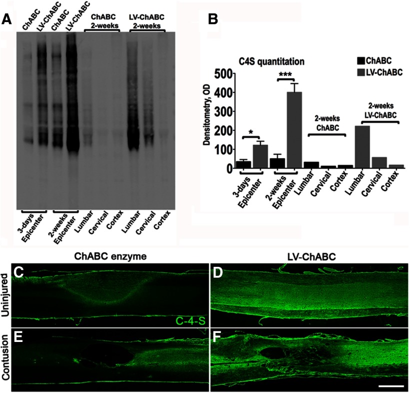
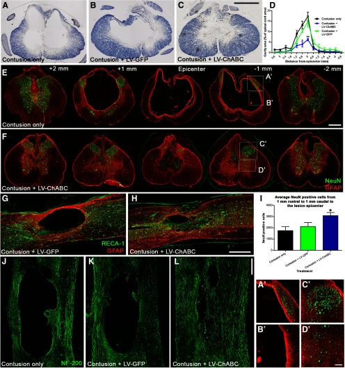
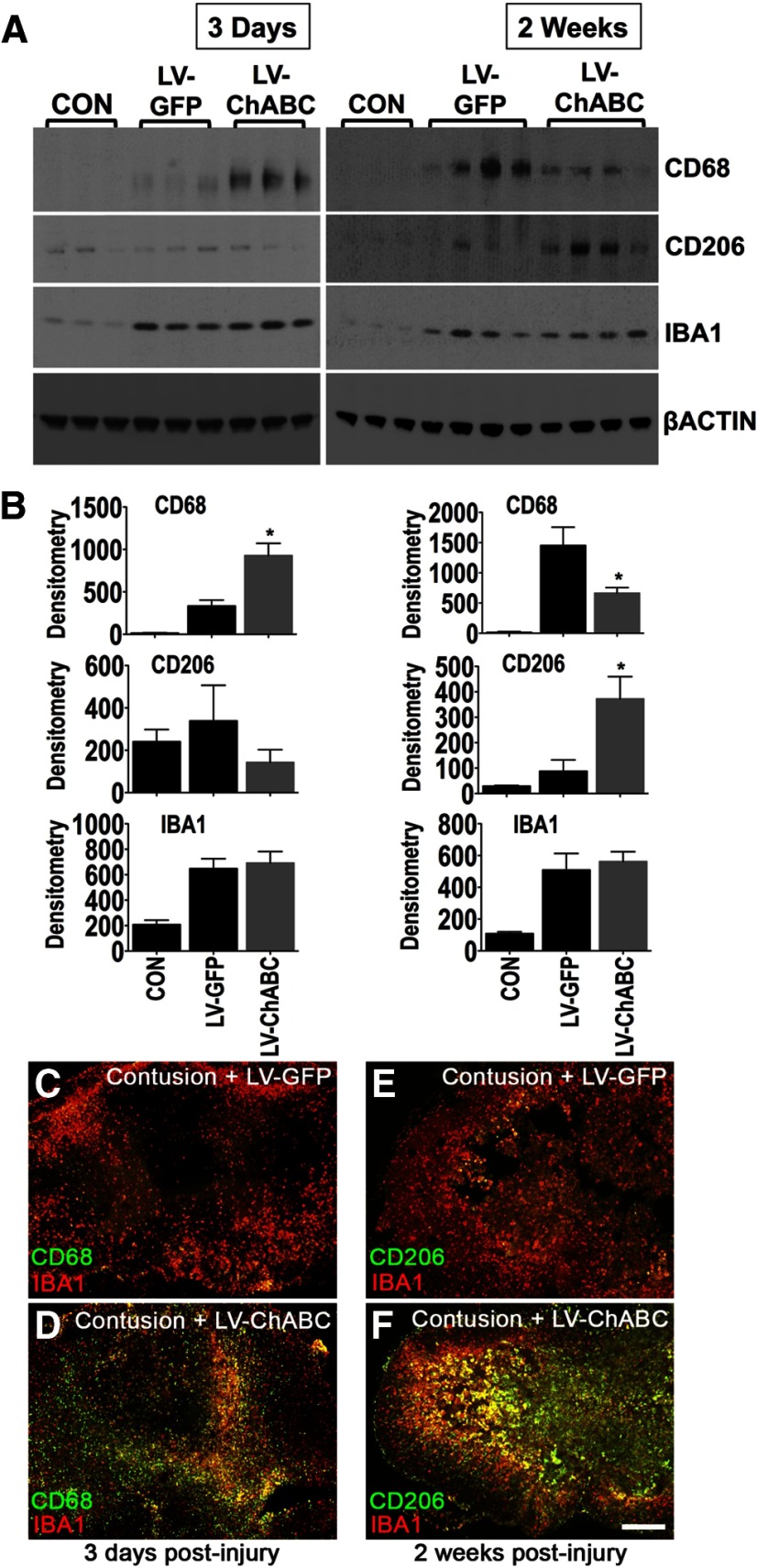
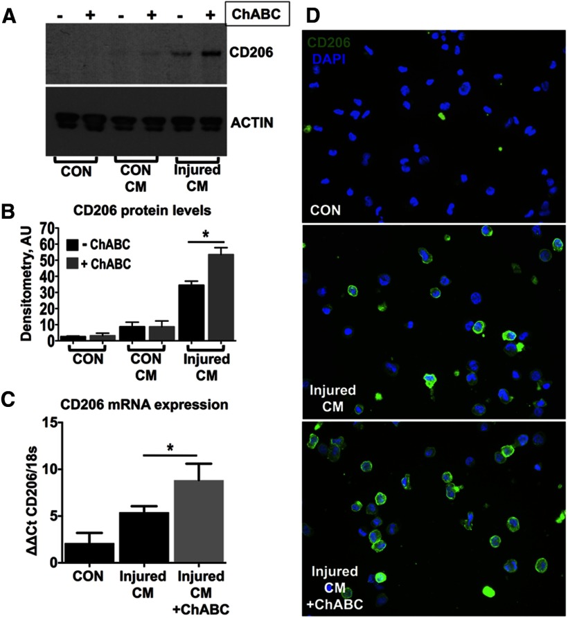
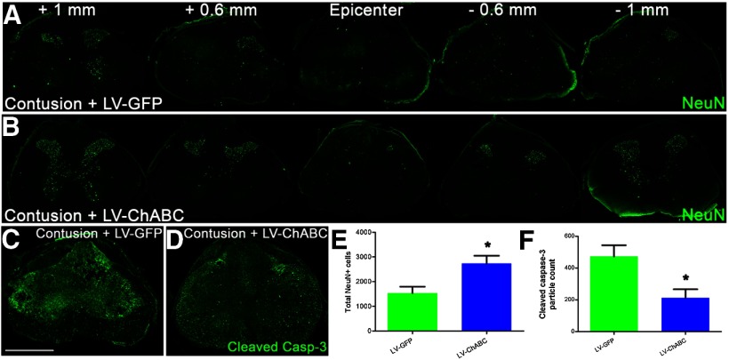
Chondroitin sulfate proteoglycans (CSPGs) inhibit repair following spinal cord injury. Here we use mammalian-compatible engineered chondroitinase ABC (ChABC) delivered via lentiviral vector (LV-ChABC) to explore the consequences of large-scale CSPG digestion for spinal cord repair. We demonstrate significantly reduced secondary injury pathology in adult rats following spinal contusion injury and LV-ChABC treatment, with reduced cavitation and enhanced preservation of spinal neurons and axons at 12 weeks postinjury, compared with control (LV-GFP)-treated animals. To understand these neuroprotective effects, we investigated early inflammatory changes following LV-ChABC treatment. Increased expression of the phagocytic macrophage marker CD68 at 3 d postinjury was followed by increased CD206 expression at 2 weeks, indicating that large-scale CSPG digestion can alter macrophage phenotype to favor alternatively activated M2 macrophages. Accordingly, ChABC treatment in vitro induced a significant increase in CD206 expression in unpolarized monocytes stimulated with conditioned medium from spinal-injured tissue explants. LV-ChABC also promoted the remodelling of specific CSPGs as well as enhanced vascularity, which was closely associated with CD206-positive macrophages. Neuroprotective effects of LV-ChABC corresponded with improved sensorimotor function, evident as early as 1 week postinjury, a time point when increased neuronal survival correlated with reduced apoptosis. Improved function was maintained into chronic injury stages, where improved axonal conduction and increased serotonergic innervation were also observed. Thus, we demonstrate that ChABC gene therapy can modulate secondary injury processes, with neuroprotective effects that lead to long-term improved functional outcome and reveal novel mechanistic evidence that modulation of macrophage phenotype may underlie these effects.

Keywords: chondroitinase; contusion; gene therapy; neuroprotection; spinal cord injury.


Figures

Figure 1.
Figure 1.
LV delivery of mammalian-compatible engineered ChABC under a PGK promoter leads to large-scale CSPG digestion in the adult mammalian spinal cord; comparison of bacterial ChABC enzyme versus LV-ChABC. A, Immunoblotting for C-4-S stubs was used to compare GAG digestion in the injury epicenter (T10) of conventional ChABC enzyme-treated and LV-ChABC-treated cords at 3 d and 2 weeks following spinal contusion. Lumbar (L2) and cervical (C4) segments as well as brain cortex were also blotted to visualize the spread of GAG digestion at 2 weeks postinjury. B, For ChABC and LV-ChABC injury epicenter comparisons, results are mean density values + SEM; n = 3 cords; *p < 0.05; ***p < 0.001, one-way ANOVA and Bonferroni's multiple-comparison test. For the lumbar, cervical, and cortex, each lane is a pool of extracts derived from three animals. C–F, Immunohistochemistry in sagittal sections through the uninjured (C, D) or the injured (E, F) thoracic spinal cord showing the C-4-S (green) digestion pattern at 8 weeks postinjection with or without contusion injury. In uninjured spinal cords, weak C-4-S immunoreactivity restricted to the injection zone was apparent 8 weeks following injection of bacterial ChABC (C), compared with intense and widespread C-4-S immunoreactivity with LV-ChABC injection (D). Abundant CSPG digestion was also observed at the injury epicenter 8 weeks following contusion and LV-ChABC injection, and the lesion site appeared to be associated with numerous tissue bridges (F). By comparison, the injury epicenter following intraspinal injection of conventional ChABC enzyme contained lower levels of digested CSPGs at 8 weeks postinjury and the epicenter had developed into a large cavity (E). These data suggest that large-scale CSPG digestion achieved by LV-ChABC may have a neuroprotective effect. Scale bar: (in F) C–F, 1 mm.
Figure 2.
Figure 2.
Gene delivery of ChABC leads to improved injury pathology and neuroprotection following spinal contusion. A–C, Eriochrome cyanine histochemistry in transverse spinal cord sections 12 weeks following spinal contusion shows a marked reduction in cavity formation at the lesion epicenter following LV-ChABC treatment (C), compared with contusion only (A) or LV-GFP treatment (B). D, Quantification of cavity area reveals a significant reduction in cavity size at the epicenter following LV-ChABC treatment as well as in the rostrocaudal spread of the cavity (asterisks indicate a significant difference compared with both contusion only and contusion plus LV-GFP; p < 0.05, 2-way repeated-measures ANOVA, Bonferroni's post hoc). E, F, Merged images of GFAP (astrocytes, red) and NeuN (neurons, green) immunoreactivity throughout the rostrocaudal axis. Dramatic changes in reactive gliosis and neuronal cell survival after contusion injury and LV-ChABC treatment (F) compared with contusion-only controls (E) were observed. I, Quantification of averaged NeuN-positive cells counted over a series of sections from 1 mm rostral to 1 mm caudal to the epicenter revealed a significant preservation of spinal neurons following LV-ChABC treatment, compared with contusion only and LV-GFP treatment; *p < 0.05, one-way ANOVA, Tukey's post hoc. G, H, The much reduced lesion epicenter in LV-ChABC-treated spinal cord contains numerous blood vessels, indicated by RECA-1 immunoreactivity (green). J–L, While large cystic cavities are apparent in chronically injured spinal cords (12 weeks postinjury) that are untreated or received LV-GFP treatment, numerous NF 200-positive axons (green) are apparent coursing through the lesion epicenter in LV-ChABC-treated spinal cords. A'–D', High-magnification images of the areas indicated in E and F. Scale bars: (in C) A–C, 500 μm; (in E) E, F, 500 μm; H, 500 μm; (in L) J–L, 500 μm; (in D') A'–D', 100 μm.
Figure 3.
Figure 3.
Gene delivery of ChABC leads to alterations in macrophage markers. A, B, Immunoblotting for CD68, CD206, and IBA1 in the injury epicenter of either LV-GFP-treated or LV-ChABC-treated cords 3 d and 2 weeks following spinal contusion (samples from individual animals are shown in each lane). Uninjured, control (CON) T10 spinal cord tissue was used for comparison. Values were normalized to β-actin. Results are mean density values + SEM; n = 3–4 cords; *p < 0.05; one-way ANOVA and Bonferroni's multiple-comparison test. C–F, Double labeling of either CD68 or CD206 (green) with IBA1 (red) shows an early increase in CD68 immunostaining in the injury epicenter 3 d after contusion and LV-ChABC injection (D) followed by increased CD206 immunostaining at 2 weeks (F), compared with LV-GFP (C, E). Scale bar: (in F) C–F, 500 μm.
Figure 4.
Figure 4.
Conditioned medium from cultured spinal cord-injured explants induces CD206 expression on unpolarized THP-1 cells. A–D, Unpolarized THP-1 human monocytic cells were treated with conditioned medium (CM) derived from either control uninjured (CON CM) or 7 d injured (injured CM) T10 spinal cord segments, cultured for 24 h at 37°C, in plain DMEM or supplemented with 0.05 U ChABC enzyme, to deglycosylate cord CSPGs. A, Immunoblotting revealed that injured CM induced CD206 upregulation on THP-1 cells and this effect was enhanced by ChABC activity. B, CD206 immunoblots were quantified by densitometry. Values were normalized to β-actin. Results are mean density values + SEM; n = 3 independent experiments; *p < 0.05; one-way ANOVA and Bonferroni's multiple-comparison test. C, The upregulation of CD206 was also confirmed at the mRNA level using quantitative PCR. Relative expression was quantified using the ΔΔCt method. CD206 PCR signal was normalized to 18s (housekeeping gene). n = 3 independent experiments; *p < 0.05. One-way ANOVA and Bonferroni's multiple-comparison test. D, CD206 immunostaining (green) in cultured THP-1 cells treated with injured CM. Nuclei were counterstained with DAPI (blue).
Figure 5.
Figure 5.
CD206-positive macrophages are closely associated with improved vascularity. A, B, At 2 weeks postinjury, the numerous CD206-positive cells (green) in the injury epicenter of LV-ChABC-treated cords were closely associated with VEGF (red), compared with LV-GFP-treated cords where few CD206-positive cells were observed in the epicenter and VEGF was concentrated mainly around the lesion border (boxed areas shown at high magnification in a' and b'). LV-ChABC-treated cords were also double stained for CD206 (green) and the endothelial cell marker RECA-1 (red), revealing CD206-positive areas that were highly vascularized in the injury epicenter (C and c' high magnification) and perilesional and meningeal blood vessels which contained CD206-positive cells at sites more distal to the injury (D and d' high magnification). Scale bars: (in A) A, B, 200 μm; (in C) C, D, 200 μm.
Figure 6.
Figure 6.
Gene delivery of ChABC leads to reduced neuronal death associated with lower levels of apoptosis at 1 week postinjury. A, B, Serial images of NeuN (neurons, green) immunoreactivity throughout the rostrocaudal axis showing greater survival of spinal neurons at 1 week postinjury following LV-ChABC treatment, compared with LV-GFP treatment. C, D, Cleaved caspase-3 immunoreactivity in the lesion epicenter reveals a clear reduction in the apoptotic marker at 1 week postinjury following LV-ChABC treatment, compared with LV-GFP treatment. E, Quantification of total NeuN-positive cells counted in serial sections from 1 mm rostral to 1 mm caudal to the epicenter revealed a significant preservation of spinal neurons following LV-ChABC treatment, compared with contusion only and LV-GFP treatment; *p < 0.05, unpaired two-tailed Student's t test. F, Quantification of cleaved caspase-3 activity in the lesion epicenter revealed reduced levels of apoptosis following LV-ChABC treatment, compared with LV-GFP controls; *p < 0.05, unpaired two-tailed Student's t test. Scale bar: (in C) A–D, 500 μm.
Figure 7.
Figure 7.
Gene delivery of ChABC promotes remodelling of specific CSPGs following spinal contusion. A, Immunoblotting for aggrecan, versican, brevican, and neurocan in the injury epicenter of either LV-GFP-treated or LV-ChABC-treated cords at 3 d and 2 weeks following spinal contusion (samples from individual animals are shown in each lane). Uninjured, control (CON) T10 spinal cord tissue was used for comparison. A switch in the expression of specific CSPGs was observed, with increased aggrecan and decreased versican apparent at 2 weeks postinjury following LV-ChABC treatment, compared with LV-GFP treatment. B, Protein levels were estimated by densitometry. Results are mean density values + SEM; n = 4 cords; *p < 0.05; one-way ANOVA and Bonferroni's multiple-comparison test. Immunoblotting against the C-4-S GAG stubs demonstrates LV-ChABC enzymatic activity in the injury epicenter.
Figure 8.
Figure 8.
Gene delivery of ChABC leads to improved sensorimotor function and spinal conduction following spinal contusion. A, Impact data showing the actual force applied to individual animals was within 10% of the intended force of 150 kdyne and mean values for each group were not significantly different (n = 14 per group; p > 0.05; 1-way ANOVA), confirming that any group differences were not due to differences in the impact force during surgery. B, Treatment with LV-ChABC leads to an early and sustained improvement in sensorimotor function, as shown by a significant reduction in footslips on the horizontal ladder task at all postinjury time points compared with contusion only and contusion plus LV-GFP treatment; p < 0.01, two-way repeated-measures ANOVA, Bonferroni's post hoc). C, Following treatment with LV-ChABC, there is a significant improvement in the percentage of sensory dorsal column axons that are capable of conducting through the injury site at a chronic (10 weeks) postinjury time point (p < 0.01, 1-way ANOVA, Tukey's post hoc). D, Example traces of recordings.
Figure 9.
Figure 9.
Gene delivery of ChABC leads to increased serotonergic innervation following spinal contusion. A–D, Serotonergic (5-HT) fiber density is significantly enhanced in the ventral horn of the lumbar spinal cord following treatment with LV-ChABC (C) compared with contusion only (A) and LV-GFP (B; quantified in D; n = 4 per group; p < 0.05, 1-way ANOVA, Tukey's post hoc). E–G, C-4-S immunoreactivity is abundant in the lumbar cord of LV-ChABC-treated animals (F), but absent in both LV-GFP and contusion-only animals (E; quantified in G; n = 4 per group; p < 0.001, 1-way ANOVA, Tukey's post hoc); boxes in A and E show regions where intensity measurements were quantified. Scale bar: (in C) A–C, 250 μm; (in F) E, F, 500 μm.
Figure 10.
Figure 10.
Long-term CSPG digestion does not lead to an enhanced pain state in uninjured animals or following spinal contusion. A, B, Assessments of withdrawal thresholds to mechanical (von Frey test; A) or thermal (Hargreaves test; B) stimulation of the forepaw following injection of saline or LV-ChABC into the C5 spinal cord revealed no significant changes in pain sensitivity over a 12 week time course. C, D, Injection of formalin to elicit a pain response in uninjured animals resulted in typical biphasic licking and biting behaviors in the affected forepaw, with no significant differences observed between groups during the early or late phases of the formalin-evoked response, indicating no enhanced sensitivity to pain following long-term treatment with LV-ChABC (n = 7–8 per group; p > 0.05 2-way repeated-measures ANOVA for time course data and Student's t test for early-phase and late-phase data). E, F, Assessments of withdrawal thresholds to mechanical (von Frey test; E) or thermal (Hargreaves test; F) stimulation of the hindpaw following T10 spinal contusion and injection of LV-GFP or LV-ChABC at the injury site revealed no significant changes in pain sensitivity over a 10 week time course (n = 7–8 per group; p > 0.05 2-way repeated-measures ANOVA).

References

    1. Alilain WJ, Horn KP, Hu H, Dick TE, Silver J. Functional regeneration of respiratory pathways after spinal cord injury. Nature. 2011;475:196–200. doi: 10.1038/nature10199. - DOI - PMC - PubMed
    1. Barritt AW, Davies M, Marchand F, Hartley R, Grist J, Yip P, McMahon SB, Bradbury EJ. Chondroitinase ABC promotes sprouting of intact and injured spinal systems after spinal cord injury. J Neurosci. 2006;26:10856–10867. doi: 10.1523/JNEUROSCI.2980-06.2006. - DOI - PMC - PubMed
    1. Bartus K, James ND, Bosch KD, Bradbury EJ. Chondroitin sulphate proteoglycans: key modulators of spinal cord and brain plasticity. Exp Neurol. 2012;235:5–17. doi: 10.1016/j.expneurol.2011.08.008. - DOI - PubMed
    1. Blesch A, Conner J, Pfeifer A, Gasmi M, Ramirez A, Britton W, Alfa R, Verma I, Tuszynski MH. Regulated lentiviral NGF gene transfer controls rescue of medial septal cholinergic neurons. Mol Ther. 2005;11:916–925. doi: 10.1016/j.ymthe.2005.01.007. - DOI - PubMed
    1. Boven LA, Van Meurs M, Van Zwam M, Wierenga-Wolf A, Hintzen RQ, Boot RG, Aerts JM, Amor S, Nieuwenhuis EE, Laman JD. Myelin-laden macrophages are anti-inflammatory, consistent with foam cells in multiple sclerosis. Brain. 2006;129:517–526. - PubMed

Publication types

MeSH terms

Substances

LinkOut - more resources