. 2014 May 9;289(19):13042-53.
doi: 10.1074/jbc.M113.537811. Epub 2014 Apr 2.

The Parkinson disease-linked LRRK2 protein mutation I2020T stabilizes an active state conformation leading to increased kinase activity

Affiliations

The Parkinson disease-linked LRRK2 protein mutation I2020T stabilizes an active state conformation leading to increased kinase activity

Soumya Ray et al. J Biol Chem. .

Abstract

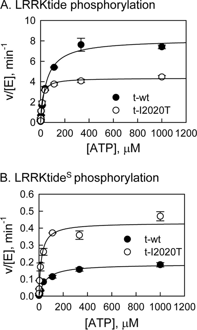
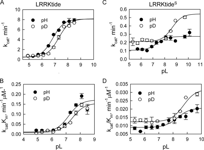
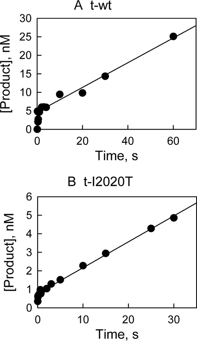
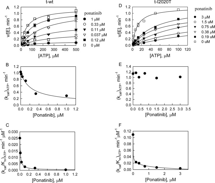
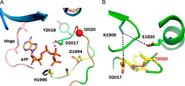
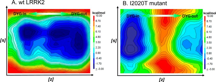
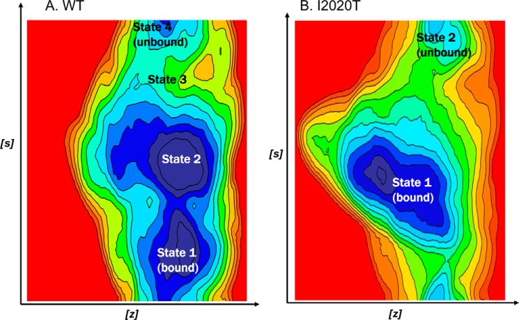
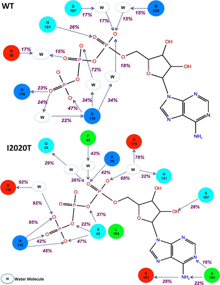
The effect of leucine-rich repeat kinase 2 (LRRK2) mutation I2020T on its kinase activity has been controversial, with both increased and decreased effects being reported. We conducted steady-state and pre-steady-state kinetic studies on LRRKtide and its analog LRRKtide(S). Their phosphorylation differs by the rate-limiting steps: product release is rate-limiting for LRRKtide and phosphoryl transfer is rate-limiting for LRRKtide(S). As a result, we observed that the I2020T mutant is more active than wild type (WT) LRRK2 for LRRKtide(S) phosphorylation, whereas it is less active than WT for LRRKtide phosphorylation. Our pre-steady-state kinetic data suggest that (i) the I2020T mutant accelerates the rates of phosphoryl transfer of both reactions by 3-7-fold; (ii) this increase is masked by a rate-limiting product release step for LRRKtide phosphorylation; and (iii) the observed lower activity of the mutant for LRRKtide phosphorylation is a consequence of its instability: the concentration of the active form of the mutant is 3-fold lower than WT. The I2020T mutant has a dramatically low KATP and therefore leads to resistance to ATP competitive inhibitors. Two well known DFG-out or type II inhibitors are also weaker toward the mutant because they inhibit the mutant in an unexpected ATP competitive mechanism. The I2020 residue lies next to the DYG motif of the activation loop of the LRRK2 kinase domain. Our modeling and metadynamic simulations suggest that the I2020T mutant stabilizes the DYG-in active conformation and creates an unusual allosteric pocket that can bind type II inhibitors but in an ATP competitive fashion.

Keywords: Enzyme Inhibitors; Enzyme Kinetics; LRRK2; Molecular modeling; Parkinson Disease.


Figures

FIGURE 1.
FIGURE 1.
Steady-state kinetic studies of LRRK2-catalyzed phosphorylation. A, phosphorylation of LRRKtide by t-WT LRRK2 (●) and the t-I2020T mutant (○) at 1 mm LRRKtide. B, phosphorylation of LRRKtideS by t-wt LRRK2 (●) and the t-I2020T mutant (○) at 2 mm LRRKtideS.
FIGURE 2.
FIGURE 2.
pL-dependent kinetic parameters for the mutant t-I2020T-catalyzed phosphorylation. A, t-I2020T-catalzyed LRRKtide phosphorylation (panels A and B). Panel A, pH (●) and pD (○) dependences of kcat with a SKIE of 1.01. Panel B, pH (●) and pD (○) dependences of kcat/Km with a SKIE of 1.1. B, t-I2020T-catalzyed LRRKtideS phosphorylation (panels C and D). Panel C, pH (●) and pD (○) dependences of kcat with a SKIE of 0.50. Panel D, pH (●) and pD (○) dependences of kcat/Km with a SKIE of 0.61.
FIGURE 3.
FIGURE 3.
Pre-steady-state kinetic studies for LRRKtide phosphorylation. The reaction was carried out at saturating concentrations of LRRKtide and ATP in the presence of 12 nm t-WT (A) and t-I2020T mutant (B). The reaction was stopped by the rapid quench flow instrument. Data were fit to a single exponential followed by a steady-state rate equation: y = A[1 − exp(−k1t)] + k2t.
FIGURE 4.
FIGURE 4.
Inhibition study of phosphorylation of LRRKtide by ponatinib. A, inhibition study of the t-WT-catalyzed phosphorylation of LRRKtide by ponatinib (panels A–C). Panel A, plot of initial velocities versus [ATP] at [ponatinib] = 1 (●), 0.3 (○), 0.1 (▾), 0.04 (▿), 0.01 (■), and 0 μm (□) all at a fixed LRRKtide concentration of 50 μm. Panels B and C, ponatinib concentration dependences of (kcat)ATP and (kcat/Km)ATP apparent values derived from analysis of the data of panel A. B, inhibition study of the t-I2020T mutant-catalyzed phosphorylation of LRRKtide by ponatinib (panels D–F). Panel D, plot of initial velocities versus [ATP] at [ponatinib] = 3 (●), 1.5 (○), 0.8 (▾), 0.4 (▿), 0.2 (■), and 0 μm (□) all at a fixed LRRKtide concentration of 50 μm. Panels E and F, ponatinib concentration dependences of (kcat)ATP and (kcat/Km)ATP apparent values derived from analysis of the data of panel D.
FIGURE 5.
FIGURE 5.
Modeling of LRRK2. A, modeling of ATP binding site of WT LRRK2. The key interacting residues and the position of Ile-2020 residue on the activation loop are highlighted. B, modeling of the I2020T mutant after MD thermalization. The side chain of I2020T participates in a network of hydrogen bond interactions that stabilizes the DYG-in conformation of the mutant.
FIGURE 6.
FIGURE 6.
FES from metadynamics simulation generated from the collective variables (s) and (z) that describe the transition of the activation loop from DYG-in to DYG-out. A, the FES of WT LRRK2 shows a contiguous low energy path (blue) that connects the two conformations forms. B, the FES of the I2020T mutant shows a deeper low energy minima corresponding to the DYG-in state and a much higher barrier separating the DYG-in and DYG-out states.
FIGURE 7.
FIGURE 7.
A, the FES of the undocking process of ATP in WT LRRK2 as a function of the angle and root mean square deviations of CVs. The FES shows an internal, partially bound and an external energy minimas. A detailed description of the various intermediate steps is provided in text. Briefly, the dissociation path of ATP involves two metastable states (states 2 and 3) with a relatively smooth transition between bound and unbound states. B, the FES of the undocking process of ATP in I2020T as a function of the angle and root mean square deviations of CVs. Unlike WT LRRK2, the dissociation appears to follow a two-step-like mechanism with a highly stable bound state that persists much longer compared with WT and then dissociate very quickly to an unbound external energy minima.
FIGURE 8.
FIGURE 8.
A, a simulation interaction diagram showing prolonged interactions between ATP, amino acid residues of WT LRRK2, and water molecules over the course of simulation. The simulation interaction diagram for WT LRRK2 shows several interactions between ATP phosphate groups and water molecules. This indicates that the ATP molecule moved fairly rapidly from the binding site to other non-native sites. The only strong persistent interaction (47%) is observed between the γ-phosphate of ATP and Arg-110 (structurally equivalent to R2026 in native LRRK2 sequence). B, a simulation interaction diagram showing prolonged interactions between ATP, amino acid residues of I2020T mutant, and water molecules over the course of simulation. Unlike WT LRRK2, the simulation interaction diagram for I2020T shows direct interaction between the ATP molecule and protein side chains that make up the hinge region and the ATP binding cavity. This indicates that the molecule has higher residence time in the ATP binding site compared with WT. In addition to the strong interaction with Arg-110 (structurally equivalent to Arg-2026 in native LRRK2 sequence), the ATP molecule also makes strong and persistent (51% total) water-mediated hydrogen bonds with Asp-170 (structurally equivalent to Asp-2017) of the DYG motif.

References

    1. Moore D. J., West A. B., Dawson V. L., Dawson T. M. (2005) Molecular pathophysiology of Parkinson's disease. Annu. Rev. Neurosci. 28, 57–87 - PubMed
    1. Paisán-Ruíz C., Jain S., Evans E. W., Gilks W. P., Simón J., van der Brug M., López de Munain A., Aparicio S., Gil A. M., Khan N., Johnson J., Martinez J. R., Nicholl D., Carrera I. M., Pena A. S., de Silva R., Lees A., Martí-Massó J. F., Pérez-Tur J., Wood N. W., Singleton A. B. (2004) Cloning of the gene containing mutations that cause PARK8-linked Parkinson's disease. Neuron 44, 595–600 - PubMed
    1. Zimprich A., Biskup S., Leitner P., Lichtner P., Farrer M., Lincoln S., Kachergus J., Hulihan M., Uitti R. J., Calne D. B., Stoessl A. J., Pfeiffer R. F., Patenge N., Carbajal I. C., Vieregge P., Asmus F., Müller-Myhsok B., Dickson D. W., Meitinger T., Strom T. M., Wszolek Z. K., Gasser T. (2004) Mutations in LRRK2 cause autosomal-dominant parkinsonism with pleomorphic pathology. Neuron 44, 601–607 - PubMed
    1. Berg D., Schweitzer K. J., Leitner P., Zimprich A., Lichtner P., Belcredi P., Brüssel T., Schulte C., Maass S., Nägele T., Wszolek Z. K., Gasser T. (2005) Type and frequency of mutations in the LRRK2 gene in familial and sporadic Parkinson's disease. Brain 128, 3000–3011 - PubMed
    1. Khan N. L., Jain S., Lynch J. M., Pavese N., Abou-Sleiman P., Holton J. L., Healy D. G., Gilks W. P., Sweeney M. G., Ganguly M., Gibbons V., Gandhi S., Vaughan J., Eunson L. H., Katzenschlager R., Gayton J., Lennox G., Revesz T., Nicholl D., Bhatia K. P., Quinn N., Brooks D., Lees A. J., Davis M. B., Piccini P., Singleton A. B., Wood N. W. (2005) Mutations in the gene LRRK2 encoding dardarin (PARK8) cause familial Parkinson's disease: clinical, pathological, olfactory and functional imaging and genetic data. Brain 128, 2786–2796 - PubMed

Publication types

MeSH terms

Substances