Review
. 2014 Mar;33(1):231-69.
doi: 10.1007/s10555-014-9498-0.

Platelets and cancer: a casual or causal relationship: revisited

Affiliations
Review

Platelets and cancer: a casual or causal relationship: revisited

David G Menter et al. Cancer Metastasis Rev. 2014 Mar.

Abstract

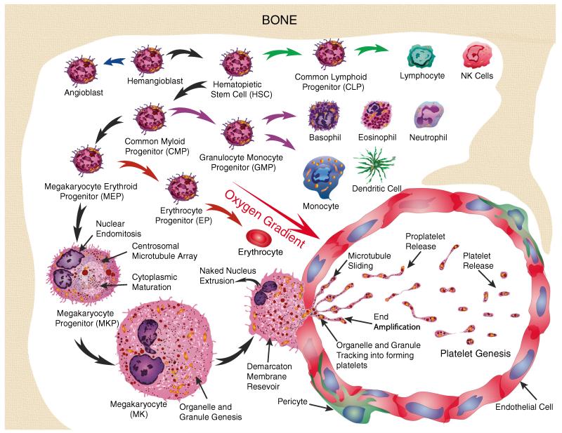
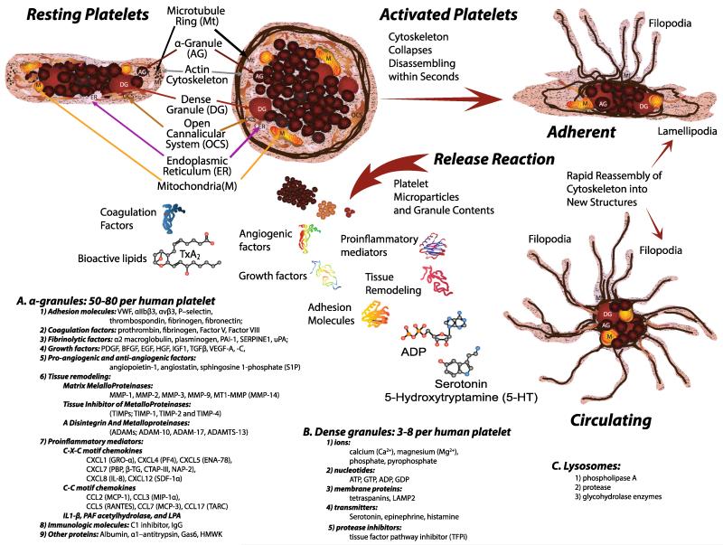
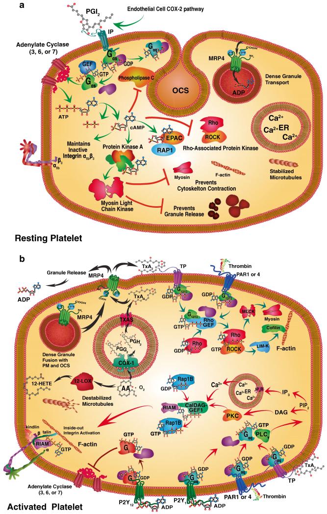
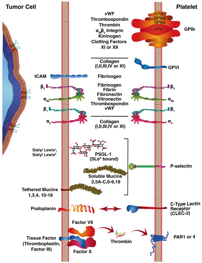
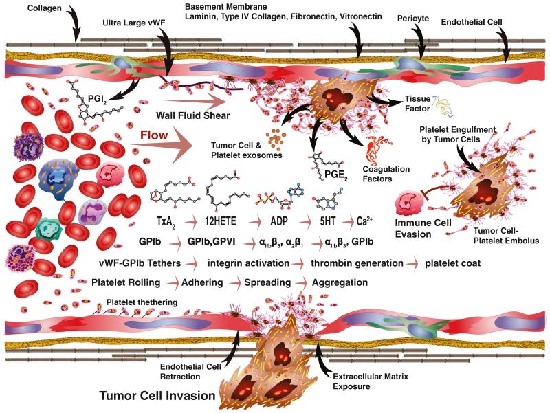
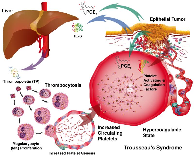
Human platelets arise as subcellular fragments of megakaryocytes in bone marrow. The physiologic demand, presence of disease such as cancer, or drug effects can regulate the production circulating platelets. Platelet biology is essential to hemostasis, vascular integrity, angiogenesis, inflammation, innate immunity, wound healing, and cancer biology. The most critical biological platelet response is serving as "First Responders" during the wounding process. The exposure of extracellular matrix proteins and intracellular components occurs after wounding. Numerous platelet receptors recognize matrix proteins that trigger platelet activation, adhesion, aggregation, and stabilization. Once activated, platelets change shape and degranulate to release growth factors and bioactive lipids into the blood stream. This cyclic process recruits and aggregates platelets along with thrombogenesis. This process facilitates wound closure or can recognize circulating pathologic bodies. Cancer cell entry into the blood stream triggers platelet-mediated recognition and is amplified by cell surface receptors, cellular products, extracellular factors, and immune cells. In some cases, these interactions suppress immune recognition and elimination of cancer cells or promote arrest at the endothelium, or entrapment in the microvasculature, and survival. This supports survival and spread of cancer cells and the establishment of secondary lesions to serve as important targets for prevention and therapy.


Figures

Fig. 1
Fig. 1
Platelet genesis occurs in the bone marrow. Hemangioblasts initially undergo divergence to form two primary lineages of cells, either angioblasts or hematopoietic stem cells (HSC). HSC subsequently form common lymphoid progenitor (CLP) or common myeloid progenitor (CMP) cells. CLP give rise to lymphocytes and other lymphoid lineage cell types while CMP generate myeloid cell types. Granulocyte monocyte progenitor (GMP) lineages include basophils, eosinophils, neutrophils, monocytes, and dendritic cells. In contrast, megakaryocyte erythroid progenitors (MEP) give rise to megakaryocytes and erythroid cells. Megakaryocyte progenitor (MKP) cell progression involves nuclear endomitosis leading to polyploidy to as high as 128n. These changes are accompanied by centrosomal microtubule array formation and cytoplasmic maturation. The increase in DNA, cytoplasm, granule, and organelle formation significantly increases the size of megakaryocytes (MK) up to 65 μm in diameter in preparation for platelet genesis. MK migrates along an oxygen gradient and send platelet-generating processes into the lumen of bone marrow capillaries that fill along sliding microtubule tracks with organelles and granules. Platelets are formed by cytoplasmic end amplification followed by proplatelet release and maturation
Fig. 2
Fig. 2
Resting platelets maintain their discoid shape using a structural ring of microtubules (Mt) and actin cytoskeleton. The plasma membrane is connected to an internal membrane reservoir called the open canalicular system (OCS). Platelets also contain organelles including endoplasmic reticulum (ER), and mitochondria (M). Resting platelets carry stores of bioactive contents in a granules (a), dense granules (b), and lysosomes (c) that are released following activation. Once activated, the platelet cytoskeleton collapses followed by extensive shape change depending upon the trigger stimulus. Lamellipodia are formed when in contact with flat surfaces such as extracellular matrix, and facilitate platelet migration. Filopodia that promote contact with other platelets or cells are formed by adherent and circulating platelets in suspension
Fig. 3
Fig. 3
Specific signaling pathways maintain the resting platelet state (a) or initiate platelet activation (b). Prostacyclin (PGI2) is primarily produced via the COX-2 pathway by endothelial cells and binds to stimulatory Gαs-coupled and GTP-GDP exchange factor (GEF) proteins through prostacyclin receptors (IP, green arrows). The resting platelet state (a) is maintained by IP-mediated Gαs-protein coupled stimulation through the synthesis of cyclic-AMP (cAMP) by adenylate cyclase (AC 3, 6, or 7). cAMP elicits a variety of effects including: inhibition of phospholipase Cγ (PLCγ) and Rho/Rho-associated kinase (ROCK) pathway. cAMP also directly stimulates exchange protein directly activated by cAMP (EPAC) and Ras-related protein 1 (Rap1) signaling, along with PKA. Protein kinase A, in turn, phosphorylates cAMP myosin light chain kinase (MLCK) that maintains an inactive actin cytoskeleton and prevents granule release as well as maintaining integrins in an inactive state. Resting platelets also accumulate ADP in dense granules via granule membrane multidrug resistance protein 4 (MRP4). Calcium stores and microtubules also remain stabilized in resting platelets. Activating platelets by contrast can involve a variety of different signaling pathways depending on the stimulus (b). Thromboxane A2 is a potent activator of platelets through the G12/13 protein coupled thromboxane receptor (TP; teal arrows). Likewise, thrombin activates protease-activated receptors (PAR) 1 and 4 through G12/13 coupled proteins. Both G12/13 coupled receptors activate Rho/ROCK kinase activity leading to MLCK phosphorylation and thereby myosin activation, and LIM domain kinase (LIM-K) that phosphorylates cofilin, which triggers actin filament disassembly. These cytoskeletal changes are accompanied by the destabilization of microtubules. TP and PAR1,4 along with ADP activation of the purinergic G-protein coupled receptor (P2Y) act upon Gaq proteins (red arrows). This activity stimulates PLCγ and the release of phosphatidylinositol 4,5-bisphophonate (PIP2) and activation of IP3 in combination with diacyl-glycerol (DAG). These events trigger calcium release and protein kinase C (PKC) activation. Ca2+ and diacylglycerol-regulated guanine nucleotide exchange factor I (CalDAG-GEF-I) catalyzes GDP for GTP exchange in Rap1B. These events activate Rap1-GTP-interacting adapter molecule (RIAM) that stimulates talin and kindlin interactions between integrin β subunits and F-actin and integrin activation. A different inhibitory Gi protein coupled receptor, P12Y, is activated by ADP and inhibits AC 3, 6, or 7. ADP is released from platelet-dense granule fusion with the plasma membrane or OCS MRP4 that transports TXA2 and ADP. These activation events initiate the synthesis of 12-hydroxyeicosatetraenoic acid (12(S)-HETE) or TXA2 from arachidonic acid (AA). TXA2 is synthesized after conversion of AA and oxygen (O2) by cyclooxygenase 1 (COX-1) to prostaglandin G2 and then H2 (PGG2, PGH2) followed by the final conversion by thromboxane A synthase (TXAS)
Fig. 4
Fig. 4
Tumor cells interact with platelets through a variety of receptors. Interactions between tumor cells and platelets can involve diverse types of cell–cell and extracellular matrix (ECM) proteins as intermediaries. Tumor cell invasion into the blood stream can expose extracellular matrix or trigger the expression of ultralarge von Willebrand factor (ULVWF) that trigger platelet rolling through tethering to collagen and other ECM proteins or endothelial cells. Platelet GPIb complexes recognize vWF tethers, along with thrombospondin, thrombin, αMβ2 integrin, kininogen, and clotting factors XI or XII. The GPIb complex consists of two sets of GPIX, GPIbβ, and GPIbα along with a centrally situated GPV protein. Subsequent stabilization of adhesive contacts with collagen exposed by tumor cells is mediated by GPVI. Tumor cell interactions with fibrinogen can also occur through intracellular adhesion molecule-1 (ICAM-1). Integrin heterodimers mediate numerous interactions with a variety of ECM proteins. The αIIbβ3 integrin is the most promiscuous and binds to fibrinogen, fibrin, fibronectin, vitronectin, thrombospondin, and vWF. Interactions with a variety of collagen molecules are mediated by α2β1. Interactions between tumor cells and cell surface carbohydrates can involve selectins. P-selectin binds to Sialyl Lewisx or Sialyl LewisxA; these interactions can also involve either tethered or soluble mucins. Similarly, carbohydrate interactions can involve C-type LECtin receptor-2 (CLEC-2) on platelets and podoplanin on tumor cells. Tissue factor expression by tumor cells can bind coagulation factor VII or X and trigger thrombin generation that activates PAR 1 or 4
Fig. 5
Fig. 5
Tumor cell interactions with platelets and other circulating factors under fluid flow are complex. Fluid shear increases from the center of blood vessels toward the vessel walls. Prostacyclin (PGI2) produced by endothelial cells inhibits platelet activation. Various molecules produced by tumor cells or other sources can activate platelets as part of a cascade of events. Progressive stimulation by TxA2-12(S)-HETE-ADP-5HT and calcium release are events that fall within the small molecule cascade. The formation of vWF-GpIb tethers support cell rolling or ultralarge vWF molecule formation, attracts platelets, and promotes their binding. Along the same cascade, GPIb-GPVI begin stabilizing adhesion and triggering αIIbβ3 and α2β1 along with other activation followed by spreading, aggregation, and invasion. Within the same cascade, thrombin generation promotes platelet coat formation and embolus formation that can enable tumor cells to evade cell-mediated immunity. Tumor cell products include exosomes, PGE2, tissue factor, and coagulation factors that act as triggers for platelet activation. Endothelial cell retraction associated with tumor cell invasion exposes those basement membrane components such as laminin, type IV collagen, fibronectin, and vitronectin involved in the tumor cell–platelet interaction cascade
Fig. 6
Fig. 6
Trousseau’s syndrome is characterized by a hypercoagulable state. The production of IL6 by tumors stimulates the production of thrombopoietin by the liver. Thrombopoietin (THPO), in turn, stimulates thrombocytosis. The production of PGE2 by tumors also heightens the responsiveness of platelets. Platelet-activating and thrombin-stimulating factors produced by tumors also elevate the potential for thrombosis. The combination of increased numbers of circulating platelets along with circulating prothrombogenic factors creates the hypercoagulable state first described by Trousseau
Fig. 7
Fig. 7
Sequence of events involved in tumor cell–platelet interactions during the arrest and extravasation of tumor cells. a The initial arrest often involves close contact between the plasma membranes of endothelial cell–platelets–tumor cells. Resting platelets (RP) associate with inactive endothelial cells (En) that are maintained in conjunction with pericytes (PC) while platelets are activated by interaction with tumor cells or their by-products in circulating plasma (Pl). b Tumor cell–platelet contacts initiate extensive platelet activation (AP) and aggregation with formation of tumor cell cytoplasmic extensions into platelet thrombus and uptake of platelet fragments by tumor cells. These interactions encompass the entire lumen of microvessels to the exclusion of other cells such as red blood cells (RBC). Platelet degranulation initially occurs in platelets closely associated with tumor cell surfaces. c Platelet degranulation becomes more extensive accompanied by additional platelet fragment engulfment by tumor cells and by endothelial cell retraction as tumor cells attach to subendothelial matrix and begin matrix degradation. d Platelet aggregates begin to disappear possibly by additional tumor cell uptake of platelet fragments. e Tumor cells begin to proliferate in capillaries. f Additional tumor cell proliferation occurs in conjunction with basement membrane (BM) degradation and invasion into the surrounding lung alveoli (Al)
Fig. 8
Fig. 8
Electron micrographs of early events in platelet–tumor cell interactions. a Elutriated W256 tumor cells in the plateau phase interacting with platelets in vitro at 10 min. Asterisked cells have lost surface microvilli (scanning electron micrograph; magnification, ×3,900). b Elutriated W256 tumor cells beginning shallow process formation (arrows) into platelets at midphase, and c deeper into platelet emboli at plateau phase (arrows) with engulfment of platelet fragments (open arrows) (transmission electron micrographs; magnifications—b ×3,000 and c ×4,300). d Interaction with and process penetration (arrows) of Lewis lung tumor cells into C57Bl/6 platelets 2 min post tail-vein injection, and e engulfing platelet fragments (arrows) at the plateau phase (transmission electron micrographs; magnifications—d ×6,800 and e ×12,000, scale bars 1 μm). ac Reprinted from [59] with permission from S. Karger AG, Basel; d, e courtesy of Menter

References

    1. Honn KV, Tang DG, Crissman JD. Platelets and cancer metastasis: a causal relationship? Cancer Metastasis Reviews. 1992;11(3-4):325–351. - PubMed
    1. Rados C. Beyond bloodletting: FDA gives leeches a medical makeover. FDA Consumer. 2004;38(5):9. - PubMed
    1. Winkel R, Tajsic N, Husum H, Schlageter M, Hanebuth G, Hoffmann R. Saphenous perforator flap. Operative Orthopädie und Traumatologie. 2013;25(2):152–161. - PubMed
    1. Kaushansky K. Historical review: megakaryopoiesis and thrombopoiesis. Blood. 2008;111(3):981–986. - PMC - PubMed
    1. Steinhubl SR. Historical observations on the discovery of platelets, platelet function testing and the first antiplatelet agent. Current Drug Targets. 2011;12(12):1792–1804. - PubMed

Publication types

LinkOut - more resources