

Volumetric texture features from higher-order images for diagnosis of colon lesions via CT colonography
- PMID: 24696313
- PMCID: PMC4185018
- DOI: 10.1007/s11548-014-0991-2
Volumetric texture features from higher-order images for diagnosis of colon lesions via CT colonography
Abstract
Purpose: Differentiation of colon lesions according to underlying pathology, e.g., neoplastic and non-neoplastic lesions, is of fundamental importance for patient management. Image intensity-based textural features have been recognized as useful biomarker for the differentiation task. In this paper, we introduce texture features from higher-order images, i.e., gradient and curvature images, beyond the intensity image, for that task.
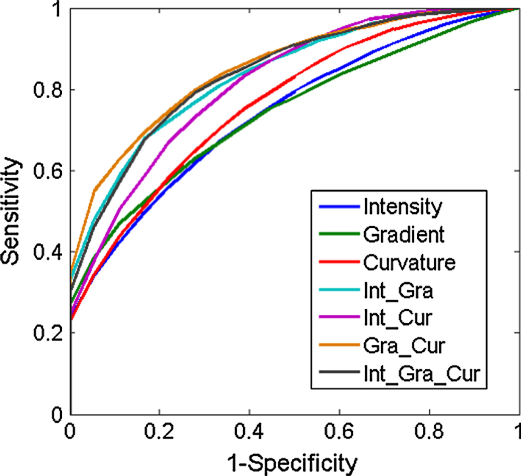
Methods: Based on the Haralick texture analysis method, we introduce a virtual pathological model to explore the utility of texture features from high-order differentiations, i.e., gradient and curvature, of the image intensity distribution. The texture features were validated on a database consisting of 148 colon lesions, of which 35 are non-neoplastic lesions, using the support vector machine classifier and the merit of area under the curve (AUC) of the receiver operating characteristics.
Results: The AUC of classification was improved from 0.74 (using the image intensity alone) to 0.85 (by also considering the gradient and curvature images) in differentiating the neoplastic lesions from non-neoplastic ones, e.g., hyperplastic polyps from tubular adenomas, tubulovillous adenomas and adenocarcinomas.
Conclusions: The experimental results demonstrated that texture features from higher-order images can significantly improve the classification accuracy in pathological differentiation of colorectal lesions. The gain in differentiation capability shall increase the potential of computed tomography colonography for colorectal cancer screening by not only detecting polyps but also classifying them for optimal polyp management for the best outcome in personalized medicine.
Keywords: CT colonography; Colorectal lesions; Computer-aided diagnosis; Curvature; Gradient; Textural biomarker; Texture feature.
Conflict of interest statement
Figures


References
-
- American Cancer Society. Cancer facts & figures 2012. Atlanta, GA: American Cancer Society; 2012.
-
- Eddy D. Screening for colorectal cancer. Ann Intern Med. 1990;113:373–384. - PubMed
-
- Gluecker T, Johnson C, Harmsen W, Offord K, Harris A, Wilson L, Ahlquist D. Colorectal cancer screening with CT colonography, colonoscopy, and double-contrast barium enema examination: prospective assessment of patient perceptions and preferences. Radiology. 2003;227(2):378–384. - PubMed
-
- Summers R, Beaulieu C, Pusanik L, Malley J, Jeffrey R, Glazer D, Napel S. Automated polyp detector for CT colonography: feasibility study. Radiology. 2000;216(1):284–290. - PubMed
-
- Yoshida H, Nappi J. Three-dimensional computer-aided diagnosis scheme for detection of colonic polyps. IEEE Trans Med Imaging. 2001;20(12):1261–1274. - PubMed
Publication types
MeSH terms
Grants and funding
LinkOut - more resources
Full Text Sources
Other Literature Sources
