. 2014 Jun;88(12):6702-13.
doi: 10.1128/JVI.00300-14. Epub 2014 Apr 2.

Characterizing functional domains for TIM-mediated enveloped virus entry

Affiliations

Characterizing functional domains for TIM-mediated enveloped virus entry

Sven Moller-Tank et al. J Virol. 2014 Jun.

Abstract

T-cell immunoglobulin and mucin domain 1 (TIM-1) and other TIM family members were recently identified as phosphatidylserine (PtdSer)-mediated virus entry-enhancing receptors (PVEERs). These proteins enhance entry of Ebola virus (EBOV) and other viruses by binding PtdSer on the viral envelope, concentrating virus on the cell surface, and promoting subsequent internalization. The PtdSer-binding activity of the immunoglobulin-like variable (IgV) domain is essential for both virus binding and internalization by TIM-1. However, TIM-3, whose IgV domain also binds PtdSer, does not effectively enhance virus entry, indicating that other domains of TIM proteins are functionally important. Here, we investigate the domains supporting enhancement of enveloped virus entry, thereby defining the features necessary for a functional PVEER. Using a variety of chimeras and deletion mutants, we found that in addition to a functional PtdSer-binding domain PVEERs require a stalk domain of sufficient length, containing sequences that promote an extended structure. Neither the cytoplasmic nor the transmembrane domain of TIM-1 is essential for enhancing virus entry, provided the protein is still plasma membrane bound. Based on these defined characteristics, we generated a mimic lacking TIM sequences and composed of annexin V, the mucin-like domain of α-dystroglycan, and a glycophosphatidylinositol anchor that functioned as a PVEER to enhance transduction of virions displaying Ebola, Chikungunya, Ross River, or Sindbis virus glycoproteins. This identification of the key features necessary for PtdSer-mediated enhancement of virus entry provides a basis for more effective recognition of unknown PVEERs.

Importance: T-cell immunoglobulin and mucin domain 1 (TIM-1) and other TIM family members are recently identified phosphatidylserine (PtdSer)-mediated virus entry-enhancing receptors (PVEERs). These proteins enhance virus entry by binding the phospholipid, PtdSer, present on the viral membrane. While it is known that the PtdSer binding is essential for the PVEER function of TIM-1, TIM-3 shares this binding activity but does not enhance virus entry. No comprehensive studies have been done to characterize the other domains of TIM-1. In this study, using a variety of chimeric proteins and deletion mutants, we define the features necessary for a functional PVEER. With these features in mind, we generated a TIM-1 mimic using functionally similar domains from other proteins. This mimic, like TIM-1, effectively enhanced transduction. These studies provide insight into the key features necessary for PVEERs and will allow for more effective identification of unknown PVEERs.


Figures

FIG 1
FIG 1
Cytoplasmic and transmembrane domains are nonessential for PVEER activity of TIM proteins. (A) Surface expression of human TIM-1, TIM-3, and TIM-4 and murine TIM-1 in HEK 293T cells. At 48 h after transfection, HEK 293T cells were incubated with polyclonal antisera against the appropriate TIM family member. Cell transfected with the empty vector were stained with anti-TIM-1 polyclonal antisera. (B, D to F, and I) Transduction of VSV virions pseudotyped with EBOV GP or LASV GPC into cells expressing TIM family members (B), IgV domain chimeras (D), cytoplasmic tail switch (E) or deletion (F) mutants, or GPI-anchored TIMs (I). Schematics of WT and chimeric proteins are shown below respective transduction data. IgV domains are represented by a crescent, MLDs are represented by a rectangle, and transmembrane/cytoplasmic domains are represented by a triangle. (C, G, and H) Surface expression of IgV domain chimeras (C), TIM cytoplasmic deletion mutants (G), and GPI-anchored TIMs (H) in HEK 293T cells at 48 h after transfection. (C) IgV chimeras were detected using polyclonal antisera specific to the WT proteins from which either the IgV domain or MLD were derived. (G) Comparison of expression of TIM family members (filled gray line) and cytoplasmic tail deletions (solid black line). (H) Surface expression of WT and GPI-anchored TIM family members with (dotted black line) or without (solid black line) PI-PLC treatment. A filled gray line represents background antisera binding. Transductions were normalized to transduction of WT TIM-1 (D and E) or equivalent WT TIM (F and I). Cells were transduced with an MOI of 0.03 (B, D, F, and I) or 0.01 (E) based on titers determined in empty vector HEK 293T cells. The data shown are the means ± the standard errors of the mean (SEM) of at least three replicates. For panels D to F and panel I, the significance was calculated using a one-sample t test comparison to 100 (normalized value for EBOV transduction in WT transfected cells (**, P < 0.001; *, P < 0.01).
FIG 2
FIG 2
Length of the MLD affects binding of EBOV virions. (A) CLUSTAL V alignment of a region of TIM-1 and mutant MLD amino acid sequences (aa 128 to 250 of WT TIM-1). Alignment was determined using MegAlign (DNASTAR). Mutants were named according to whether they retained amino-terminal (N), middle (M), or carboxy-terminal (C) sequences of the MLD and the lengths of their MLDs. (B) Binding of FITC-labeled VSV virions pseudotyped with EBOV GP to HEK 293T cells expressing WT TIM-1 or MLD mutants as assessed by flow cytometry. All mutants shown retain the carboxy-terminal sequence of the MLD. Representative histograms are shown with black lines representing virus binding and gray-filled lines representing background fluorescence. (C) Representative contour plots of FITC-labeled virion binding versus TIM-1 expression. Transfected cells were stained with MLD specific, MAb AKG7, after binding virus. (D) Dot plot of virus binding to TIM-1 versus estimated mucin length. The data were quantified from binding studies in panel B by using mean fluorescence intensity (MFI) of virus-positive cells. (E) Immunoblot demonstrating equivalent amounts of HA-tagged TIM protein after normalization of HEK 293T supernatants. (F) Binding of soluble WT TIM-1 and MLD mutants to PtdSer liposomes. Serial dilutions of normalized TIM-1 protein were incubated with ELISA plates prebound overnight with PtdSer liposomes (50 μM). The data are means ± the SEM of at least three replicates.
FIG 3
FIG 3
EBOV transduction correlates with TIM-1 MLD length. (A to C) Transduction of MLD mutant or WT TIM-1-transfected HEK 293T cells with VSV pseudotyped with EBOV GP (A and B) or LASV GPC (C). Transduction relative to WT TIM-1 is shown as a bar graph (A) or plotted against mucin length (B and C). Trend lines were fitted for panels B and C with a one-site specific binding nonlinear regression with Hill Slope and linear regression, respectively, using Prism (GraphPad). Cells were transduced with an MOI of 0.01 as titered in empty-vector HEK 293T cells. (D) Virus binding (MFI) from Fig. 2D plotted against the percent transduction data. (E) Transduction of pseudovirions into cells expressing a single MLD mutant (●) or various mixtures of MLD mutants (○). Transduction for mixed MLD populations is plotted using the average MLD lengths of the MLD mutants transfected. Transduction data for panels A, B, and D are normalized to TIM-1 expression as assessed by surface staining (% TIM-positive cells), and all data are means ± the SEM of at least three replicates. For panel A, significance was calculated by using a one-sample t test comparison to 100 (**, P < 0.001).
FIG 4
FIG 4
Domains of TIM-1 required for PVEER activity can be functionally substituted. (A) The amino acid sequences of TIM-1 MLD switch chimeras are aligned with the MLD of TIM-1 (aa 128 to 298) (CLUSTAL V). Terminal amino acids of the aligned sequences are noted. (B, D, and F) Transduction of transfected HEK 293T cells with VSV virions pseudotyped with viral glycoprotein as noted in each panel. Schematics of chimeras are shown below transduction data (B and D) or adjacent to expression data (E). (B) Transduction of cells expressing TIM/αDG chimeras. The MLD of murine αDG replaced or added in addition to TIM-1 MLD generated αDG-MLD or 2×MLD constructs, respectively. (C) Representative histograms of TIM-1/PRR and TIM-1/RAGE chimeras (black line) and WT TIM-1 (filled gray line) surface expression as determined using ARD5. (D) Transduction of cells expressing TIM-1/PRR and TIM-1/RAGE chimeras compared to transduction into cells expressing equivalent amounts of wild-type TIM-1 IgV domain. Empty vector transduction data are compared to the same TIM-1 transduction data as h1RAGE and h1RAGEh1cyto since these had the lowest expression levels. (E) Expression of WT TIM-1 and AnxV-αDG-GPI as detected using TIM-1 IgV and MLD specific MAbs ARD5 and AKG7 and AnxV antisera (black line) compared to background IgG binding (filled gray). (F) Transduction of cells expressing either TIM-1 or AnxV-αDG-GPI. Cells were transduced with an MOI of 0.02 (EBOV and LASV in panels B and F) or 0.03 (EBOV and LASV in panel D and all other viruses in panel F) as titered in empty vector transfected HEK 293T cells. The data are shown as means ± the SEM of at least three replicates. Significance was calculated using a two-sample t test comparison to empty vector (B) or a one-sample t test comparison to 100 (D) (**, P < 0.001).
FIG 5
FIG 5
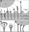
Model of MLD role in PVEER-mediated entry. (A) Accessibility of PtdSer on the surface of virions to IgV domain of TIMs is dependent on length of MLD. Short MLDs or structured stalks cannot project the IgV domain above the majority of cell surface proteins. Mid-length MLDs in some cases are blocked by adjacent proteins, but in other cases are partially accessible if the local environment is shorter. Full-length MLDs, however, are fully accessible and project the IgV above the majority of cell surface proteins. MLDs containing structured regions in addition to extended conformations or with additional length function similarly. (B) Potential flexibility of the MLD provides a larger area of interaction for longer MLD proteins than shorter MLD proteins. (C) Flexibility of the MLD may also allow for TIMs to conform to curvature of virions and promote binding of multiple TIMs. Crescent shapes represent PtdSer-binding domains, ovals represent the IgC domains from RAGE or similar structured domains, and coils represent serine/threonine/proline-rich beta-turn helices.

References

    1. Feigelstock D, Thompson P, Mattoo P, Zhang Y, Kaplan GG. 1998. The human homolog of HAVcr-1 codes for a hepatitis A virus cellular receptor. J. Virol. 72:6621–6628 - PMC - PubMed
    1. Jemielity S, Wang JJ, Chan YK, Ahmed AA, Li W, Monahan S, Bu X, Farzan M, Freeman GJ, Umetsu DT, Dekruyff RH, Choe H. 2013. TIM-family proteins promote infection of multiple enveloped viruses through virion-associated phosphatidylserine. PLoS Pathog. 9:e1003232. 10.1371/journal.ppat.1003232 - DOI - PMC - PubMed
    1. Meertens L, Carnec X, Lecoin MP, Ramdasi R, Guivel-Benhassine F, Lew E, Lemke G, Schwartz O, Amara A. 2012. The TIM and TAM families of phosphatidylserine receptors mediate dengue virus entry. Cell Host Microbe 12:544–557. 10.1016/j.chom.2012.08.009 - DOI - PMC - PubMed
    1. Kondratowicz AS, Lennemann NJ, Sinn PL, Davey RA, Hunt CL, Moller-Tank S, Meyerholz DK, Rennert P, Mullins RF, Brindley M, Sandersfeld LM, Quinn K, Weller M, McCray PB, Jr, Chiorini J, Maury W. 2011. T-cell immunoglobulin and mucin domain 1 (TIM-1) is a receptor for Zaire Ebolavirus and Lake Victoria Marburgvirus. Proc. Natl. Acad. Sci. U. S. A. 108:8426–8431. 10.1073/pnas.1019030108 - DOI - PMC - PubMed
    1. Moller-Tank S, Kondratowicz AS, Davey RA, Rennert PD, Maury W. 2013. Role of the phosphatidylserine receptor TIM-1 in enveloped-virus entry. J. Virol. 87:8327–8341. 10.1128/JVI.01025-13 - DOI - PMC - PubMed

Publication types

MeSH terms