

Hilar cholangiocarcinoma: diagnosis, treatment options, and management
- PMID: 24696835
- PMCID: PMC3955000
- DOI: 10.3978/j.issn.2304-3881.2014.02.05
Hilar cholangiocarcinoma: diagnosis, treatment options, and management
Abstract
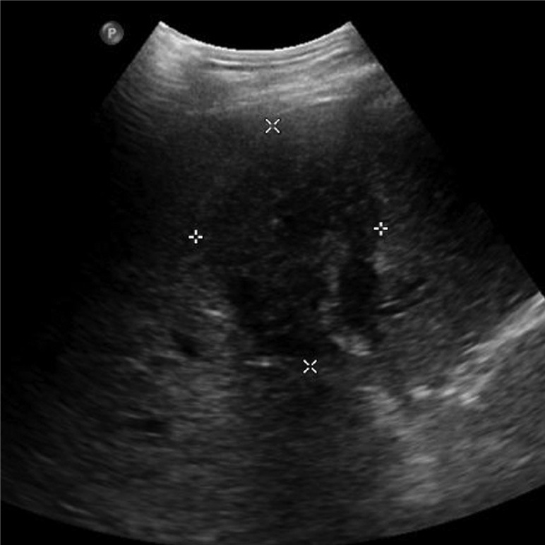
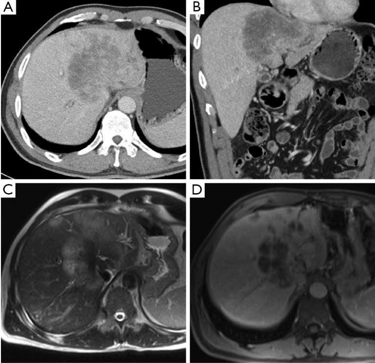
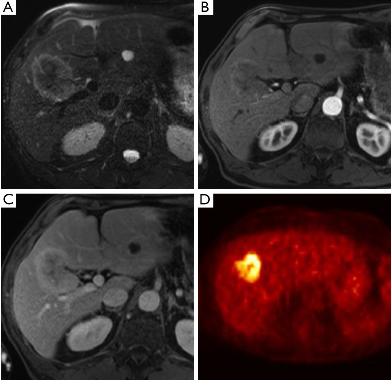
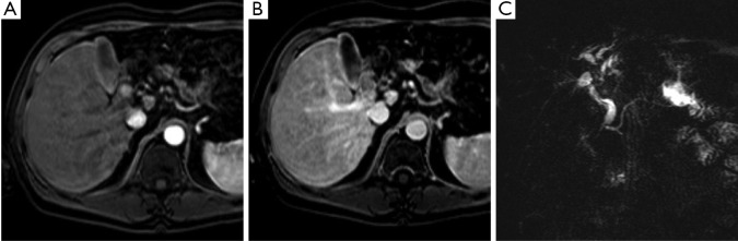
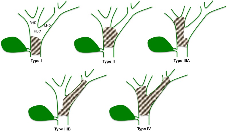
Hilar cholangiocarcinoma (HC) is a rare disease with a poor prognosis which typically presents in the 6(th) decade of life. Of the 3,000 cases seen annually in the United States, less than one half of these tumors are resectable. A variety of risk factors have been associated with HC, most notably primary sclerosing cholangitis (PSC), biliary stone disease and parasitic liver disease. Patients typically present with abdominal pain, pruritis, weight loss, and jaundice. Computed topography (CT), magnetic resonance imaging (MRI), and ultrasound (US) are used to characterize biliary lesions. Endoscopic retrograde cholangiopancreatography (ERCP) and percutaneous transhepatic cholangiography (PTC) assess local ductal extent of the tumor while allowing for therapeutic biliary drainage. MRCP has demonstrated similar efficacies to PTC and ERCP in identifying anatomic extension of tumors with less complications. Treatment consists of surgery, radiation, chemotherapy and photodynamic therapy. Biliary drainage of the future liver remnant should be performed to decrease bilirubin levels thereby facilitating future liver hypertrophy. Standard therapy consists of surgical margin-negative (R0) resection with extrahepatic bile duct resection, hepatectomy and en bloc lymphadenectomy. Local resection should not be undertaken. Lymph node invasion, tumor grade and negative margins are important prognostic indicators. In instances where curative resection is not possible, liver transplantation has demonstrated acceptable outcomes in highly selected patients. Despite the limited data, chemotherapy is indicated for patients with unresectable tumors and adequate functional status. Five-year survival after surgical resection of HC ranges from 10% to 40% however, recurrence can be as high as 50-70% even after R0 resection. Due to the complexity of this disease, a multi-disciplinary approach with multimodal treatment is recommended for this complex disease.
Keywords: Cholangiocarcinoma (CC); biliary neoplasm; hilar.
Figures
References
-
- Altemeier WA, Gall EA, Zinninger MM, et al. Sclerosing carcinoma of the major intrahepatic bile ducts. AMA Arch Surg 1957;75:450-60; discussion 460-1 - PubMed
-
- Klatskin G.Adenocarcinoma of the Hepatic Duct at Its Bifurcation within the Porta Hepatis. An Unusual Tumor with Distinctive Clinical and Pathological Features. Am J Med 1965;38:241-56 - PubMed
Publication types
LinkOut - more resources
Full Text Sources
Research Materials