. 2014 Jun;224(6):624-33.
doi: 10.1111/joa.12174. Epub 2014 Apr 3.

A multi-scale structural study of the porcine anterior cruciate ligament tibial enthesis

Affiliations

A multi-scale structural study of the porcine anterior cruciate ligament tibial enthesis

Lei Zhao et al. J Anat. 2014 Jun.

Abstract

Like the human anterior cruciate ligament (ACL), the porcine ACL also has a double bundle structure and several biomechanical studies using this model have been carried out to show the differential effect of these two bundles on macro-level knee joint function. It is hypothesised that if the different bundles of the porcine ACL are mechanically distinct in function, then a multi-scale anatomical characterisation of their individual enthesis will also reveal significant differences in structure between the bundles. Twenty-two porcine knee joints were cleared of their musculature to expose the intact ACL following which ligament-bone samples were obtained. The samples were fixed in formalin followed by decalcification with formic acid. Thin sections containing the ligament insertion into the tibia were then obtained by cryosectioning and analysed using differential interference contrast (DIC) optical microscopy and scanning electron microscopy (SEM). At the micro-level, the anteromedial (AM) bundle insertion at the tibia displayed a significant deep-rooted interdigitation into bone, while for the posterolateral (PL) bundle the fibre insertions were less distributed and more focal. Three sub-types of enthesis were identified in the ACL and related to (i) bundle type, (ii) positional aspect within the insertion, and (iii) specific bundle function. At the nano-level the fibrils of the AM bundle were significantly larger than those in the PL bundle. The modes by which the AM and PL fibrils merged with the bone matrix fibrils were significantly different. A biomechanical interpretation of the data suggests that the porcine ACL enthesis is a specialized, functionally graded structural continuum, adapted at the micro-to-nano scales to serve joint function at the macro level.

Keywords: anterior cruciate ligament; enthesis; functional adaptation; macro-, micro-, and nano-level structure.


Figures

Figure 1
Figure 1
Frontal view of the left knee dissected and the femur rotated (flexed). The bundles are teased apart to show their distinct and separate gross morphologies to reveal the anteromedial (AM) bundle, and its insertion into the anterior aspect of the tibia, and the posterolateral (PL) bundle.
Figure 2
Figure 2
(A) ACL-on-bone block showing the fanning out of the ligament fibres (black arrows) at the tibial end of the ligament–bone insertion. (B) Cross-sectional view of the ACL showing the ‘crescent’ shaped wrapping (dotted line) of the AM bundle about the PL bundle. The arrowhead in (B) indicates the anterior aspect. AM, anteromedial; PL, posterolateral.
Figure 3
Figure 3
Sagittal view of the AM bundle insertion showing the intense interdigitation of ligament fibres with its rigid substrate (see the boxed region). The vertical white line indicates the depth of fibre rooting. Optical image was obtained by top illumination of the fully hydrated section.
Figure 4
Figure 4
Medial-most sagittal views of (A) the AM bundle and (B) the PL bundle showing the strongly oblique orientation of the cement line (solid line) with respect to the approximate plane of the tibial plateau (see dotted lines). The general direction of the ligament fibre alignment (see arrow) is at a relatively high angle to the cement line.
Figure 5
Figure 5
Lateral-most sagittal views of (A) the AM bundle and (B) the PL bundle showing the irregular profile of the cement line (solid line) with respect to the approximate plane of the tibial plateau (see dotted lines). The AM bundle in (A) shows a sharp turn of the ligament fibres (black arrow) into the fibrocartilage–bone substrate, and also deep fibre rooting (vertical line). Conversely the PL bundle in (B) shows a significantly reduced depth of ligament fibre rooting (see vertical line).
Figure 6
Figure 6
(A) Sagittal view of the AM bundle of the ACL showing fibrous rooting with a significant degree of deep interdigitation. (B) Sagittal view of the PL bundle of the ACL showing relatively less depth of insertion into the bone. Boxed regions in (A) and (B) are enlarged in (C) and (D), respectively. The cement lines are shown with dotted lines.
Figure 7
Figure 7
Sagittal view of posterior aspect of (A) the AM bundle and (B) the PL bundle at the same magnification and showing different degrees of mineralization at the ligament–bone junction. There are three visible zones in the AM bundle, but four in the PL bundle.
Figure 8
Figure 8
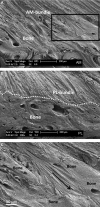
SEM images of (A) AM bundle and (B) PL bundle insertions into bone. The interdigitation of ligament fibres into the bony substrate (e.g. see boxed region in A enlarged in C) of the AM bundle insertion contrasts with that of the PL bundle. The PL bundle insertion (dotted line in B) shows a distinct and near-linear boundary between ligament and bone.
Figure 9
Figure 9
SEM of AM bundle showing fibrillar-level integration with bone. Note also the presence of near-transversely organised collagen fibrils forming well-defined ‘nodal’ clusters along the ligament–bone interface (see arrows).
Figure 10
Figure 10
SEM image of PL bundle insertion showing how the ligamentous fibrils end in a shallow bone socket.
Figure 11
Figure 11
SEM images taken at the same magnification showing fibril diameter differences in (A) the AM bundle, (B) the PL bundle, and (C) the bone.
Figure 12
Figure 12
Schematics summarising the important structure–function features of the ACL enthesis as shown in the present study. (A) Schematic illustrating how the ACL is designed to resist both anterior tibial translation and internal tibial rotation. The crescent-like shape of the AM bundle about the PL may help to distribute shear forces between these two functionally different ligament bundles. (B) One of the insertion types seen in the medial-most section is that of an adaptive bone contour that results in an orthogonal ligament alignment with regard to the cement line. (C). Another type of insertion, more common in the PL bundle, is the functionally graded material change from soft-to-hard tissue, which would appear to be designed to resist torsional or distortional/twisting strains. (D) The deep-rooted fibres, seen mainly in the lateral aspect of the ACL and most significantly in the AM bundle insertion, would appear to be designed to resist multiply directed pull-out forces.

Similar articles

Cited by

References

    1. Amis AA. The functions of the fibre bundles of the anterior cruciate ligament in anterior drawer, rotational laxity and the pivot shift. Knee Surg Sports Traumatol Arthrosc. 2012;20:613–620. - PubMed
    1. Amis AA, Dawkins GP. Functional anatomy of the anterior cruciate ligament. Fibre bundle actions related to ligament replacements and injuries. J Bone Joint Surg Br. 1991a;73:260–267. - PubMed
    1. Amis AA, Dawkins GPC. Functional anatomy of the anterior cruciate ligament. J Bone Joint Surg. 1991b;73-B:8. - PubMed
    1. Benjamin M, Evans EJ, Copp L. The histology of tendon attachments to bone in man. J Anat. 1986;149:89–100. - PMC - PubMed
    1. Benjamin M, Kumai T, Milz S, et al. The skeletal attachment of tendons – tendon ‘entheses’. Comp Biochem Physiol A Mol Integr Physiol. 2002;133:931–945. - PubMed

Publication types

LinkOut - more resources