. 2015 Jan;172(2):583-92.
doi: 10.1111/bph.12712. Epub 2014 Jul 1.

Does PKC activation increase the homologous desensitization of μ opioid receptors?

Affiliations

Does PKC activation increase the homologous desensitization of μ opioid receptors?

Seksiri Arttamangkul et al. Br J Pharmacol. 2015 Jan.

Abstract

Background and purpose: This study examined the role of agents known to activate PKC on morphine-induced desensitization of μ-opioid receptors (MOP receptors) in brain slices containing locus coeruleus neurons.

Experimental approach: Intracellular recordings were obtained from rat locus coeruleus neurons. Two measurements were used to characterize desensitization, the decline in hyperpolarization induced by application of a saturating concentration of agonist (acute desensitization) and the decrease in hyperpolarization induced by a subsaturating concentration of [Met](5) enkephalin (ME) following washout of the saturating concentration (sustained desensitization). Internalization of MOP receptors was studied in brain slices prepared from transgenic mice expressing Flag-MOP receptors. The subcellular distribution of activated PKC was examined using a novel fluorescent sensor of PKC in HEK293 cells.

Key results: The phorbol esters (PMA and PDBu) and muscarine increased acute desensitization induced by a saturating concentration of morphine and ME. These effects were not sensitive to staurosporine. Staurosporine did not block the decline in hyperpolarization induced by muscarine. PDBu and muscarine did not affect sustained desensitization induced by ME nor did phorbol esters or muscarine change the trafficking of MOP receptors induced by morphine or ME. The distribution of activated PKC measured in HEK293 cells differed depending on which phorbol ester was applied.

Conclusions and implications: This study demonstrates a distinct difference in two measurements that are often used to evaluate desensitization. The measure of decline correlated well with the reduction in peak amplitudes caused by PKC activators implicating the modification of other factors rather than MOP receptors.

Linked articles: This article is part of a themed section on Opioids: New Pathways to Functional Selectivity. To view the other articles in this section visit http://dx.doi.org/10.1111/bph.2015.172.issue-2.

Keywords: PKC; desensitization; fluorescent PKC sensor; hyperpolarization; internalization; muscarine; phorbol esters; staurosporine; μ opioid receptor.


Figures

Figure 1
Figure 1
PKC activators increased the acute decline from peak hyperpolarization in the continuous presence of morphine (MP). (A) Intracellular recording of membrane potential during 10 min morphine application in the presence of PKC activator, control (left), PDBu (0.2 μM, middle) and muscarine (10 μM, right). (B) The decline in hyperpolarization normalized to the peak hyperpolarization (*P < 0.05; ***P < 0.001).
Figure 2
Figure 2
Representative recording in which [Met5]-enkephalin (ME; 0.3 μM) and noradrenaline (NA; 100 μM plus 1 μM cocaine to block the NA transporter) were tested before and after applications of (A) PDBu (0.2 μM), (B) muscarine (10 μM), or (C) staurosporine (1 μM, pre-incubated for 60 min) plus muscarine (10 μM). The hatched bars indicate 10 min of cropped recording. (D) The decline in response showed an inverse linear correlation with peak amplitudes. The linear regression analysis gave a best-fit value with R2 = 0.8473, slope = −1.163 ± 0.2468, y-intercept = 41.03 ± 5.54 and x-intercept = 35.28. (E) Summary showing the reduction in hyperpolarization induced by ME (300 nM) after the application of the PKC activators. Analyses using Student's paired, two-tailed t-test showed that the hyperpolarizations in the presence of PDBu (1 μM), PMA (1 μM), PDBu (0.2 μM) and muscarine (10 μM) were significantly different from their untreated controls (##P < 0.01; #P < 0.05). (F) Summary of the acute decline in hyperpolarization after the treatment of slices with staurosporine (1 μM, over 60 min) and then co-application with either PDBu (0.2 μM) or muscarine (10 μM). ns = not significantly different.
Figure 3
Figure 3
The effect of PKC activators on ME-induced MOP receptor desensitization. Representative recordings measuring acute desensitization, sustained desensitization, and recovery under (A) control conditions and (B) in the presence of muscarine (10 μM, pre-incubation for 10 min and in all solutions). ME (300 nM) was applied before and 5, 15, 30 and 45 min following desensitization (ME 30 μM, 10 min). (C) Summary showing the acute decline from peak hyperpolarization in the presence of PKC activators: PMA (1 μM), PDBu (1 μM) and muscarine (10 μM), *P < 0.01. (D) Sustained desensitization/recovery measured as the relative hyperpolarization activated by ME (300 nM) at 5, 15, 30 and 45 min post-desensitization (ME 30 μM, 10 min) normalized to the initial ME-induced hyperpolarization (300 nM, prepulse).
Figure 4
Figure 4
Opioid-induced Flag-MOP receptor (MOPr) internalization after PKC activation (A) Flag-MOP receptors in LC neurons labelled with M1-A594 (first column) in live brain slices, perfused with PKC activators 10 min (second column), then morphine (15 μM) or ME (30 μM) 10 min (third column) in the presence of PKC activators and calcium-free ACSF containing 0.5 mM EGTA 10 min (last column). (B) Summary of % receptor internalization.
Figure 5
Figure 5
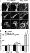
PMA-induced translocation of PKC to the plasma membrane. (A) A fluorescent PKC sensor (upward-DAG) was imaged in Flag-MOP receptor (MOPr) expressing HEK293 cells. Confocal images were taken before (left column) or following 10 min exposure (middle column) to either morphine (15 μM, top), PDBu (0.2 μM, middle) or PMA (1 μM, bottom). Flag-MOP receptors were labelled with M1-Alexa 594 (right column) to identify the plasma membrane and to confirm that Flag-MOP receptors were expressed in individual cells. (B) Summary of upward-DAG fluorescence intensities at the membrane and cytosol and the ratio of membrane/cytosol expressed as % of upward-DAG fluorescence measured before drug treatment. *P < 0.05; **P < 0.01, ***P < 0.001, paired t-test. Scale bar = 20 μm.

References

    1. Alexander SPH, Benson HE, Faccenda E, Pawson AJ, Sharman JL, Spedding M, et al. CGTP Collaborators. The Concise Guide to PHARMACOLOGY 2013/14: G protein-coupled receptors. Br J Pharmacol. 2013a;170:1459–1581. - PMC - PubMed
    1. Alexander SPH, Benson HE, Faccenda E, Pawson AJ, Sharman JL, Spedding M, et al. CGTP Collaborators. The Concise Guide to PHARMACOLOGY 2013/14: Ion channels. Br J Pharmacol. 2013b;170:1607–1651. - PMC - PubMed
    1. Arttamangkul S, Quillinan N, Low MJ, von Zastrow M, Pintar J, Williams JT. Differential activation and trafficking of μ-opioid receptors in brain slices. Mol Pharmacol. 2008;74:972–979. - PMC - PubMed
    1. Arttamangkul S, Lau EK, Lu HW, Williams JT. Desensitization and trafficking of μ-opioid receptor in locus coeruleus neurons: modulation by kinases. Mol Pharm. 2012;81:348–355. - PMC - PubMed
    1. Bailey CP, Kelly E, Henderson G. Protein kinase C activation enhances morphine-induced rapid desensitization of mu-opioid receptors in mature rat locus coeruleus neurons. Mol Pharmacol. 2004;66:1592–1598. - PubMed

Publication types

MeSH terms

LinkOut - more resources