. 2014 May 15;173(3):472-80.
doi: 10.1016/j.ijcard.2014.03.086. Epub 2014 Mar 20.

15-deoxy-Δ¹²,¹⁴-PGJ₂ promotes inflammation and apoptosis in cardiomyocytes via the DP2/MAPK/TNFα axis

Affiliations

15-deoxy-Δ¹²,¹⁴-PGJ₂ promotes inflammation and apoptosis in cardiomyocytes via the DP2/MAPK/TNFα axis

Chintan N Koyani et al. Int J Cardiol. .

Abstract

Background: Prostaglandins (PGs), lipid autacoids derived from arachidonic acid, play a pivotal role during inflammation. PGD₂ synthase is abundantly expressed in heart tissue and PGD₂ has recently been found to induce cardiomyocyte apoptosis. PGD₂ is an unstable prostanoid metabolite; therefore the objective of the present study was to elucidate whether its final dehydration product, 15-deoxy-Δ¹²,¹⁴-PGJ₂ (15d-PGJ₂, present at high levels in ischemic myocardium) might cause cardiomyocyte damage.

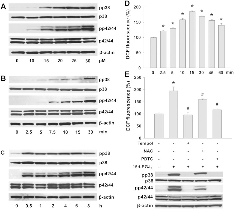
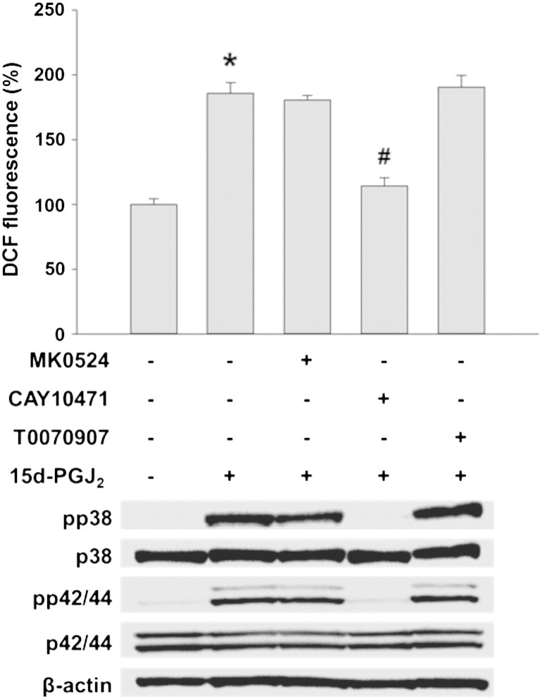
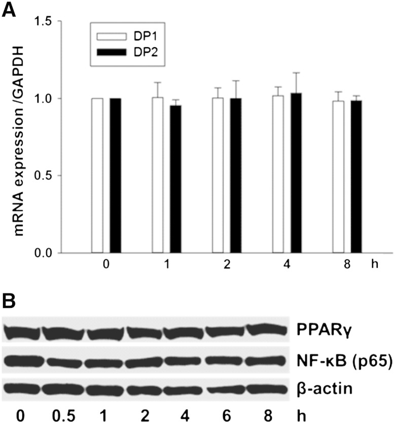
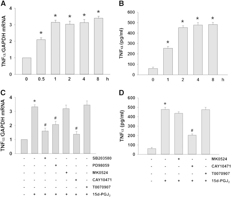
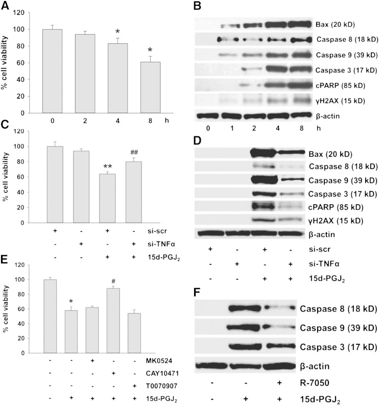
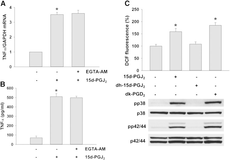
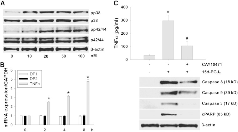
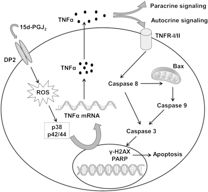
Methods and results: Using specific (ant)agonists we show that 15d-PGJ₂ induced formation of intracellular reactive oxygen species (ROS) and phosphorylation of p38 and p42/44 MAPKs via the PGD2 receptor DP2 (but not DP1 or PPARγ) in the murine atrial cardiomyocyte HL-1 cell line. Activation of the DP2-ROS-MAPK axis by 15d-PGJ₂ enhanced transcription and translation of TNFα and induced apoptosis in HL-1 cardiomyocytes. Silencing of TNFα significantly attenuated the extrinsic (caspase-8) and intrinsic apoptotic pathways (bax and caspase-9), caspase-3 activation and downstream PARP cleavage and γH2AX activation. The apoptotic machinery was unaffected by intracellular calcium, transcription factor NF-κB and its downstream target p53. Of note, 9,10-dihydro-15d-PGJ₂ (lacking the electrophilic carbon atom in the cyclopentenone ring) did not activate cellular responses. Selected experiments performed in primary murine cardiomyocytes confirmed data obtained in HL-1 cells namely that the intrinsic and extrinsic apoptotic cascades are activated via DP2/MAPK/TNFα signaling.

Conclusions: We conclude that the reactive α,β-unsaturated carbonyl group of 15d-PGJ₂ is responsible for the pronounced upregulation of TNFα promoting cardiomyocyte apoptosis. We propose that inhibition of DP2 receptors could provide a possibility to modulate 15d-PGJ₂-induced myocardial injury.

Keywords: 15d-PGJ(2); Apoptosis; Cardiomyocytes; PGD(2) receptor; TNFα.


Figures

Fig. 1
Fig. 1
15d-PGJ2 promotes MAPK activation in cardiomyocytes via intracellular redox balance. (A) HL-1 cells were treated with indicated concentrations of 15d-PGJ2 for 1 h to follow pp38 and pp42/44 MAPK expression by Western blot. Cells were incubated with 15d-PGJ2 (15 μM) for indicated time periods to follow (B/C) pp38 and pp42/44 MAPK expression and (D) ROS generation by DCF fluorescence. Cells were incubated with ROS scavengers (Tempol [1 mM], NAC [5 mM] or PDTC [1 mM]) for 30 min prior to 15d-PGJ2 treatments (15 μM) for 1 h to follow (E, upper panel) ROS generation and (E, lower panel) pp38 and pp42/44 MAPK expression. For Western blot analysis protein lysates were subjected to SDS-PAGE, proteins were transferred to nitrocellulose membranes and immunoreactive bands were visualized using specific primary and secondary antibodies. For normalization, membranes were stripped and probed with primary antibodies against β-actin as well as total p38 and p42/44 MAPK. One representative blot (A–C and E) out of three is shown. (D) For detection of intracellular ROS levels cells were incubated with carboxy-H2DCFDA (10 μM) for 30 min after treatment with 15d-PGJ2. DCF fluorescence levels of vehicle (0.1% DMSO)-treated cells were set 100% and values are expressed as mean ± SEM (n = 6). * p ≤ 0.05 vs. untreated.
Fig. 2
Fig. 2
DP2 mediates ROS production and MAPK activation in cardiomyocytes in response to 15d-PGJ2. HL-1 cells were incubated with MK0524 (100 nM), CAY10471 (1 μM) or T0070907 (20 μM) for 30 min prior to 15d-PGJ2 (15 μM) treatments for 15 min to follow ROS levels by DCF fluorescence (upper panel) and 1 h to follow pp38 and pp42/44 MAPK expression (lower panel). For detection of intracellular ROS levels cells were incubated with carboxy-H2DCFDA (10 μM) for 30 min after treatment with 15d-PGJ2. DCF fluorescence levels of vehicle (0.1% DMSO)-treated cells were set 100% and values are expressed as mean ± SEM (n = 6). * p ≤ 0.05 vs. untreated and # p ≤ 0.05 vs. 15d-PGJ2 treated. For Western blot analysis protein lysates were subjected to SDS-PAGE, proteins were transferred to nitrocellulose membranes and immunoreactive bands were visualized using specific primary and secondary antibodies. For normalization, membranes were stripped and probed with primary antibodies against β-actin as well as total p38 and p42/44 MAPK. One representative blot out of three is shown.
Fig. 3
Fig. 3
Expression of PG receptors DP1, DP2 and PPARγ. HL-1 cells were incubated with 15d-PGJ2 (15 μM) for indicated time periods to follow (A) DP1 and DP2 mRNA expression using qPCR as well as (B) PPARγ and NF-κB protein expression using Western blot. For mRNA expression cells were lysed, RNA was isolated and qPCR was performed using specific primers. Values are expressed as mean ± SEM (n = 6). For Western blot analysis protein lysates were subjected to SDS-PAGE, proteins were transferred to nitrocellulose membranes and immunoreactive bands were visualized using specific primary and secondary antibodies. For normalization, membranes were stripped and probed with primary antibodies against β-actin. One representative blot (B) out of three is shown.
Fig. 4
Fig. 4
DP2-ROS-MAPK signaling increases TNFα transcription and translation in 15d-PGJ2-stimulated cardiomyocytes. HL-1 cells were incubated with 15d-PGJ2 (15 μM) for indicated time periods to follow (A) TNFα mRNA expression by qPCR and (B) TNFα protein concentrations in the cell culture medium by ELISA. Cells were incubated with SB203580 (10 μM), PD98059 (25 μM), MK0524 (100 nM), CAY10471 (1 μM) or T0070907 (20 μM) for 30 min prior to 15d-PGJ2 (15 μM) treatment for 4 h to follow (C) TNFα mRNA expression and (D) TNFα protein concentrations in the cell culture medium. After treatment with 15d-PGJ2 the cell culture medium was removed and cells were lysed to isolate RNA for qPCR analysis using specific primers (A/C) (n = 6). The cell culture medium was used to quantitate TNFα concentrations using ELISA (B/D) (n = 3). Values are expressed as mean ± SEM. * p ≤ 0.05 vs. untreated and # p ≤ 0.05 vs. 15d-PGJ2.
Fig. 5
Fig. 5
15d-PGJ2-induced TNFα activates the apoptotic machinery in cardiomyocytes. HL-1 cells were incubated with 15d-PGJ2 (15 μM) for indicated time periods to measure (A) cell viability using MTT assay and to follow (B) apoptotic markers by Western blot. Alternatively, cells were transfected with scrambled negative control siRNA (si-scr, 40 nM) or siRNA against TNFα (si-TNFα, 40 nM) for 6 h and grown for 24 h followed by 15d-PGJ2 (15 μM) treatment for 4 h to follow (C) cell viability and (D) apoptotic markers. Cells were incubated with (E) MK0524 (100 nM), CAY10471 (1 μM), T0070907 (20 μM) or (F) R-7050 (30 μM) for 30 min prior to 15d-PGJ2 (15 μM) treatments for 4 h to follow (E) cell viability and (F) markers of apoptosis. For Western blot analysis protein lysates were subjected to SDS-PAGE, proteins were transferred to nitrocellulose membranes and immunoreactive bands were visualized using specific primary and secondary antibodies. For normalization, membranes were stripped and probed with primary antibody against β-actin. One representative blot out of three (B, D, and F) is shown. Only cleaved (but not pro-forms) of PARP (cPARP), caspase-3, -8 and -9 are shown. Cell viability was measured following incubating cells with MTT (0.5 mg/ml) for 30 min after treatment with 15d-PGJ2. Optical density levels of vehicle (0.1% DMSO)-treated cells were set 100% and values are expressed as mean ± SEM (A, C and E) (n = 6). * p ≤ 0.05 vs. untreated, # p ≤ 0.05 vs. 15d-PGJ2, ** p ≤ 0.05 vs. si-scr and ## p ≤ 0.05 vs. si-scr + 15d-PGJ2.
Fig. 6
Fig. 6
15d-PGJ2-induced signaling is calcium-independent and mediated via its cyclopentenone ring. HL-1 cells were incubated with EGTA-AM (50 μM) for 30 min prior to 15d-PGJ2 (15 μM) treatment for 4 h to follow (A) intracellular TNFα mRNA expression and (B) TNFα protein concentrations in the cell supernatant. After treatment with 15d-PGJ2 the cell culture medium was removed and cells were lysed to isolate RNA for qPCR analysis (A, n = 6) using specific primers. The cell culture medium was used for TNFα quantification by ELISA (B, n = 3). Alternatively, cells were incubated with 15d-PGJ2 (15 μM), 9,10-dihydro-15d-PGJ2 (dh-15d-PGJ2, 15 μM) or 13,14-dihydro-15-keto PGD2 (dk-PGD2, 1 μM) for 1 h to follow (C, upper panel) ROS levels by DCF fluorescence and (C, lower panel) pp38 and pp42/44 MAPK expression. For Western blot analysis protein lysates were subjected to SDS-PAGE, proteins were transferred to nitrocellulose membranes and immunoreactive bands were visualized using specific primary and secondary antibodies. For normalization, membranes were stripped and probed with primary antibodies against total p38 and p42/44 MAPK. One representative blot (C, lower panel) out of three is shown. * p ≤ 0.05 vs. untreated.
Fig. 7
Fig. 7
15d-PGJ2 induces apoptotic pathways in primary murine cardiomyocytes. (A) Cardiomyocytes were treated with indicated concentrations of 15d-PGJ2 for 1 h to follow pp38 and pp42/44 MAPK expression by Western blot. (B/C) Primary cardiomyocytes were incubated with 15d-PGJ2 (50 nM) (B) for indicated time periods to follow TNFα mRNA expression by qPCR, or (C) for 8 h to follow TNFα protein concentrations in the cell culture medium (upper panel) and markers of apoptosis (lower panel). For Western blot analysis protein lysates were subjected to SDS-PAGE, proteins were transferred to nitrocellulose membranes and immunoreactive bands were visualized using specific primary and secondary antibodies. For normalization, membranes were stripped and probed with a primary antibody against β-actin. One representative blot out of two (A and C, lower panel) is shown. Only cleaved (but not pro-forms) of PARP (cPARP), caspase-3, -8 and -9 are shown. After treatment with 15d-PGJ2 the cell culture medium was removed and cells were lysed to isolate RNA for qPCR analysis using specific primers (B) (n = 3). The cell culture medium was used for quantitation of TNFα by ELISA (C, upper panel) (n = 3). Values are expressed as mean ± SEM. * p ≤ 0.05 vs. untreated and # p ≤ 0.05 vs. 15d-PGJ2.
Fig. 8
Fig. 8
Summary of 15d-PGJ2-induced signaling events in cardiomyocytes observed during the present study.

Comment in

Similar articles

Cited by

References

    1. Agrawal R., Agrawal N., Koyani C.N., Singh R. Molecular targets and regulators of cardiac hypertrophy. Pharmacol Res. 2010;61:269–280. - PubMed
    1. Kalra B.S., Roy V. Efficacy of metabolic modulators in ischemic heart disease: an overview. J Clin Pharmacol. 2012;52:292–305. - PubMed
    1. Dumont E.A., Hofstra L., van Heerde W.L. Cardiomyocyte death induced by myocardial ischemia and reperfusion: measurement with recombinant human annexin-V in a mouse model. Circulation. 2000;102:1564–1568. - PubMed
    1. Ryu S.K., Mallat Z., Benessiano J. Phospholipase A2 enzymes, high-dose atorvastatin, and prediction of ischemic events after acute coronary syndromes. Circulation. 2012;125:757–766. - PubMed
    1. Gross G.J., Falck J.R., Gross E.R., Isbell M., Moore J., Nithipatikom K. Cytochrome P450 and arachidonic acid metabolites: role in myocardial ischemia/reperfusion injury revisited. Cardiovasc Res. 2005;68:18–25. - PubMed

Publication types

MeSH terms