

In situ visualization of plasma cells producing antibodies reactive to Porphyromonas gingivalis in periodontitis: the application of the enzyme-labeled antigen method
- PMID: 24698402
- PMCID: PMC4282379
- DOI: 10.1111/omi.12052
In situ visualization of plasma cells producing antibodies reactive to Porphyromonas gingivalis in periodontitis: the application of the enzyme-labeled antigen method
Abstract
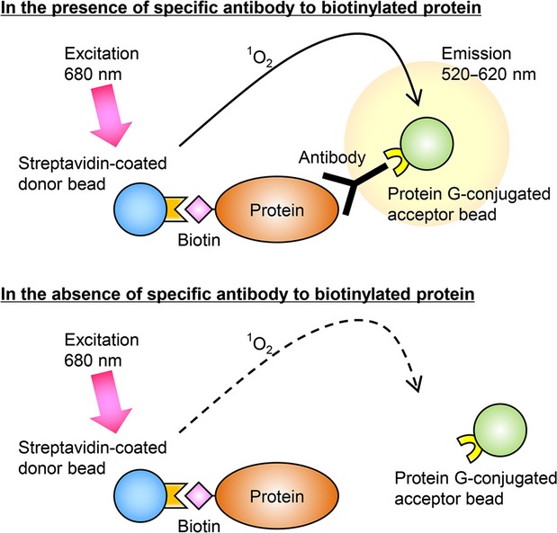
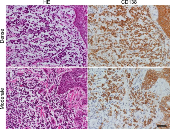
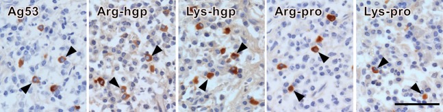
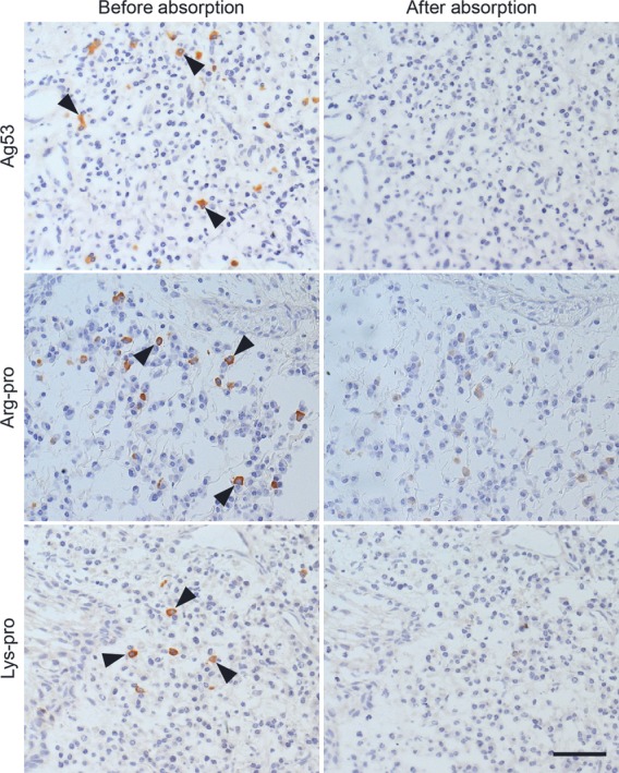
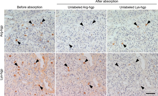
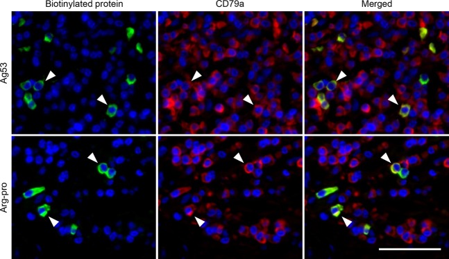
Porphyromonas gingivalis is a keystone periodontal pathogen. Histologocally, the gingival tissue in periodontitis shows dense infiltration of plasma cells. However, antigens recognized by antibodies secreted from the immunocytes remain unknown. The enzyme-labeled antigen method was applied to detecting plasma cells producing P. gingivalis-specific antibodies in biopsied gingival tissue of periodontitis. N-terminally biotinylated P. gingivalis antigens, Ag53 and four gingipain domains (Arg-pro, Arg-hgp, Lys-pro and Lys-hgp) were prepared by the cell-free protein synthesis system using wheatgerm extract. With these five labeled proteins as probes, 20 lesions of periodontitis were evaluated. With the AlphaScreen method, antibodies against any one of the five P. gingivalis antigens were detected in 11 (55%) serum samples and 17 (85%) tissue extracts. Using the enzyme-labeled antigen method on paraformaldehyde-fixed frozen sections of gingival tissue, plasma cells were labeled with any one of the five antigens in 17 (94%) of 18 specimens, in which evaluable plasma cells were detected. The positivity rates in periodontitis were significantly higher than those found previously in radicular cysts (20% in sera and 33% in tissue extracts with the AlphaScreen method, and 25% with the enzyme-labeled antigen method). Our findings directly indicate that antibodies reactive to P. gingivalis are locally produced in the gingival lesions, and that inflammatory reactions against P. gingivalis are involved in periodontitis.
Keywords: AlphaScreen method; antigen 53; gingipain; wheat germ cell free protein synthesis.
© 2014 The Authors. Molecular Oral Microbiology published by John Wiley & Sons Ltd.
Figures
Similar articles
-
Specific in situ visualization of plasma cells producing antibodies against Porphyromonas gingivalis in gingival radicular cyst: application of the enzyme-labeled antigen method.J Histochem Cytochem. 2011 Jul;59(7):673-89. doi: 10.1369/0022155411408906. Epub 2011 Apr 27. J Histochem Cytochem. 2011. PMID: 21525188 Free PMC article.
-
Serum antibody levels against Porphyromonas gingivalis extract and its chromatographic fraction in chronic and aggressive periodontitis.J Int Acad Periodontol. 2008 Apr;10(2):50-8. J Int Acad Periodontol. 2008. PMID: 18564729
-
Porphyromonas gingivalis-mediated shedding of extracellular matrix metalloproteinase inducer (EMMPRIN) by oral epithelial cells: a potential role in inflammatory periodontal disease.Microbes Infect. 2011 Dec;13(14-15):1261-9. doi: 10.1016/j.micinf.2011.07.009. Epub 2011 Jul 28. Microbes Infect. 2011. PMID: 21835259
-
Enzyme-labeled Antigen Method: Development and Application of the Novel Approach for Identifying Plasma Cells Locally Producing Disease-specific Antibodies in Inflammatory Lesions.Acta Histochem Cytochem. 2016 Feb 27;49(1):7-19. doi: 10.1267/ahc.15030. Epub 2016 Feb 26. Acta Histochem Cytochem. 2016. PMID: 27006517 Free PMC article. Review.
-
The chronicles of Porphyromonas gingivalis: the microbium, the human oral epithelium and their interplay.Microbiology (Reading). 2008 Oct;154(Pt 10):2897-2903. doi: 10.1099/mic.0.2008/021220-0. Microbiology (Reading). 2008. PMID: 18832296 Free PMC article. Review.
Cited by
-
LASP1, CERS6, and Actin Form a Ternary Complex That Promotes Cancer Cell Migration.Cancers (Basel). 2023 May 16;15(10):2781. doi: 10.3390/cancers15102781. Cancers (Basel). 2023. PMID: 37345118 Free PMC article.
-
Autoantibody Profiling Using Human Autoantigen Protein Array and AlphaScreen.Methods Mol Biol. 2024;2766:107-128. doi: 10.1007/978-1-0716-3682-4_12. Methods Mol Biol. 2024. PMID: 38270871
-
Application of an enzyme-labeled antigen method for visualizing plasma cells producing antibodies against Strep A, a carbohydrate antigen of Streptococcus pyogenes, in recurrent tonsillitis.Microbiol Immunol. 2015 Jan;59(1):13-27. doi: 10.1111/1348-0421.12213. Microbiol Immunol. 2015. PMID: 25403787 Free PMC article.
-
CEBPγ facilitates lamellipodia formation and cancer cell migration through CERS6 upregulation.Cancer Sci. 2021 Jul;112(7):2770-2780. doi: 10.1111/cas.14928. Epub 2021 May 4. Cancer Sci. 2021. PMID: 33934437 Free PMC article.
-
Differential immune cell infiltrations between healthy periodontal and chronic periodontitis tissues.BMC Oral Health. 2020 Oct 27;20(1):293. doi: 10.1186/s12903-020-01287-0. BMC Oral Health. 2020. PMID: 33109155 Free PMC article.
References
-
- Aguilera Galaviz LA, Aceves Medina Mdel C, Estrada Garcia IC. Detection of potentially cariogenic strains of Streptococcus mutans using the polymerase chain reaction. J Clin Pediatr Dent. 2002;27:47–51. - PubMed
-
- Anusaksathien O, Singh G, Matthews N, Dolby AE. Autoimmunity to collagen in adult periodontal disease: immunoglobulin classes in sera and tissue. J Periodontal Res. 1992;27:55–61. - PubMed
-
- Beikler T, Ehmke B, Wittstock M, Schmidt H, Karch H, Flemmig TF. Serum antibody reactivity against recombinant PrtC of Porphyromonas gingivalis following periodontal therapy. J Periodontal Res. 2003;38:276–281. - PubMed
-
- Berglundh T, Donati M. Aspects of adaptive host response in periodontitis. J Clin Periodontol. 2005;32(Suppl 6):87–107. - PubMed
Publication types
MeSH terms
Substances
LinkOut - more resources
Full Text Sources
Other Literature Sources
