

Scaffolds to control inflammation and facilitate dental pulp regeneration
- PMID: 24698696
- PMCID: PMC4208618
- DOI: 10.1016/j.joen.2014.01.019
Scaffolds to control inflammation and facilitate dental pulp regeneration
Abstract
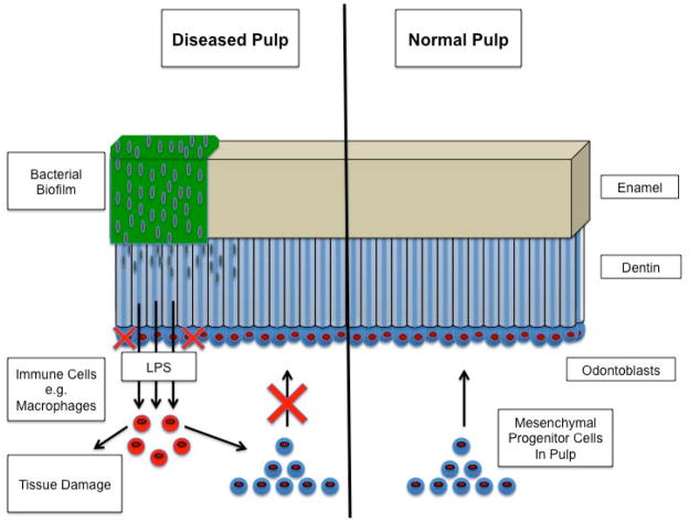
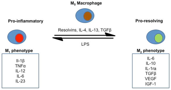
In dentistry, the maintenance of a vital dental pulp is of paramount importance because teeth devitalized by root canal treatment may become more brittle and prone to structural failure over time. Advanced carious lesions can irreversibly damage the dental pulp by propagating a sustained inflammatory response throughout the tissue. Although the inflammatory response initially drives tissue repair, sustained inflammation has an enormously destructive effect on the vital pulp, eventually leading to total necrosis of the tissue and necessitating its removal. The implications of tooth devitalization have driven significant interest in the development of bioactive materials that facilitate the regeneration of damaged pulp tissues by harnessing the capacity of the dental pulp for self-repair. In considering the process by which pulpitis drives tissue destruction, it is clear that an important step in supporting the regeneration of pulpal tissues is the attenuation of inflammation. Macrophages, key mediators of the immune response, may play a critical role in the resolution of pulpitis because of their ability to switch to a proresolution phenotype. This process can be driven by the resolvins, a family of molecules derived from fatty acids that show great promise as therapeutic agents. In this review, we outline the importance of preserving the capacity of the dental pulp to self-repair through the rapid attenuation of inflammation. Potential treatment modalities, such as shifting macrophages to a proresolving phenotype with resolvins are described, and a range of materials known to support the regeneration of dental pulp are presented.
Keywords: Biomaterials; dental pulp; endodontics; inflammation; pulp regeneration; resolvins; tissue engineering.
Copyright © 2014. Published by Elsevier Inc.
Conflict of interest statement
They declare no conflicts of interests relating to the authorship and/or publication of this article.
Figures
References
-
- Harris R, Griffin CJ. The fine structure of the mature odontoblasts and cell rich zone of the human dental pulp. Australian Dental Journal. 1969;14(3):168–177. - PubMed
-
- Berkovitz BK, Holland GR, Moxham B. Oral anatomy, embryology and histology. Mosby Edinburgh; 2002.
-
- Nanci A. Ten Cate’s oral histology: development, structure, and function. Elsevier Health Sciences; 2007.
-
- Linde A. The extracellular matrix of the dental pulp and dentin. Journal of dental research. 1985;64(Spec No):523–529. - PubMed
Publication types
MeSH terms
Substances
Grants and funding
LinkOut - more resources
Full Text Sources
Other Literature Sources
