. 2014 Jul;8(5):927-41.
doi: 10.1016/j.molonc.2014.02.005. Epub 2014 Mar 24.

Multi-marker analysis of circulating cell-free DNA toward personalized medicine for colorectal cancer

Affiliations

Multi-marker analysis of circulating cell-free DNA toward personalized medicine for colorectal cancer

Florent Mouliere et al. Mol Oncol. 2014 Jul.

Abstract

Development of a Q-PCR-based assay for the high-performance analysis of circulating cell-free DNA (ccfDNA) requires good knowledge of its structure and size. In this work, we present the first visual determination of ccfDNA by Atomic Force Microscopy (AFM) on plasma samples from colorectal cancer (CRC) patients and healthy donors. In addition to the examination of fragment size distribution profile as performed by Q-PCR, this analysis confirms that ccfDNA is highly fragmented and that more than 80% of ccfDNA fragments in CRC plasma are below 145 bp. We adapted an Allele-Specific Blocker (ASB) Q-PCR to small ccfDNA fragments to determine simultaneously the total ccfDNA concentration, the presence of point mutation, the proportion of mutated allele, and a ccfDNA integrity index. The data validated analytically these four parameters in 124 CRC clinical samples and 71 healthy individuals. The multi-marker method, termed Intplex, enables sensitive and specific non-invasive analysis of tumor ccfDNA, which has great potential in terms of cost, quality control, and easy implementation in every clinical center laboratory.

Keywords: Circulating cell-free DNA; Colorectal cancer; Multi-marker analysis; qPCR.


Figures

Figure 1
Figure 1
CcfDNA is highly fragmented. A. Size profile distribution of ccfDNA amounts in CRC (colorectal cancer) of two patients (named CRC1 and CRC2) and healthy individual plasma. The size profile distribution was evaluated with a Q‐PCR experiment and corresponding primer of targeting amplicons of increasing size. B. Proportions of ccfDNA per size range in % of the total observed ccfDNA with Q‐PCR. C. AFM visualization of ccfDNA fragments extracted and purified from stage IV CRC patient plasma. A representative visual determination of ccfDNA is shown here. D. Size profile distribution of ccfDNA fragments (n) measured with AFM experiments.
Figure 2
Figure 2
Schematic representation of Intplex primer system, localization of each primer set on a hypothetical DNA template and the oligoblocking primer. This schema is given as an illustration and an equivalent Intplex construction was carried out when we analyzed the V600E mutation of the BRAF gene.
Figure 3
Figure 3
CcfDNA concentration measurement with Intplex. A. Representation of the repeatability and reproducibility of the ccfDNA quantification results with the Intplex primer system on ccfDNA extracted with a QIAgen Blood mini kit. B. Schematic representation of the targeted KRAS and BRAF sequences in Fig. 3C, and respective localization of the primers. C. Quantification with Intplex of genomic DNA (black) and quantification of ccfDNA from a mCRC patient (grey). DNA concentration was determined with different primer sets: one specific to an intronic sequence of KRAS, two specific to two sequences on an exonic area of KRAS, and two specific to two BRAF sequences. D. Evaluation of the ccfDNA concentration with the Intplex Q‐PCR system on two different genes (KRAS and BRAF) and at different concentrations (n = 98, corresponding to CRC plasma samples).
Figure 4
Figure 4
Technical validation of the ccfDNA mutation measurement with Intplex. Q‐PCR raw amplification curve of genomic DNA from the mutant cell line containing the tested mutation (red) was serially diluted five times into high‐concentrated WT gDNA (green) from human placenta (Sigma). All the experimental points were obtained in duplicate. In this figure, only the KRAS G12A dilution is shown as a representative example of our sensitivity data.
Figure 5
Figure 5
Technical validation of the ccfDNA fragmentation measurement with Intplex. The fragmentation is calculated with a representative index, the DII (DNA Integrity Index). The DII was estimated from the ratio of the DNA concentration obtained by targeting a 300‐bp sequence and a 60‐bp sequence in a same locus. A. Evaluation of the DII with Intplex Q‐PCR system on two different genes (KRAS and BRAF). Correlation between the KRAS and BRAF DII was evaluated with a Spearman analysis (r = 0.817, p < 0.001). B. Estimation of the DII on the targeted gene (KRAS, BRAF) and the sequence within this gene (exon, intron). The denominations "conv" and "inv" refer to a particular primer system schematized in Fig. 3, B. The ccfDNA used for the DII evaluation was isolated from a mCRC patient and from a placenta DNA extract (to serve as the genomic DNA control). C. Ratio of ccfDNA DII from genomic DNA control and from CRC plasma extract.
Figure 6
Figure 6
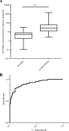
A: Comparison of the quantification of plasma ccfDNA from healthy individuals (n = 71) and mCRC patients (n = 98). The concentration observed in mCRC patients is significantly greater than those of healthy individuals (p = 0.0022). The data are expressed in ng/mL plasma. B: Diagnosis predictive capacity of total ccfDNA concentration to distinguish plasma from mCRC patients and healthy subjects. ROC Curve representation deriving from the univariate logistic analysis corresponding to the total ccfDNA (AUC = 0.91). The observed concentration of total ccfDNA from late stage mCRC patients (n = 98) was compared to the total ccfDNA concentration of healthy individuals (n = 71). This curve measures the accuracy of biomarkers displaying the relationship between sensitivity (true‐positive rate, y‐axes) and 1‐specificity (false‐positive rate, x‐axes).

Similar articles

Cited by

References

    1. Amado, R.G. , Wolf, M. , Peeters, M. , Van Cutsem, E. , Siena, S. , Freeman, D.J. , Juan, T. , Sikorski, R. , Suggs, S. , Radinsky, R. , Patterson, S.D. , Chang, D.D. , 2008. Wild-type KRAS is required for panitumumab efficacy in patients with metastatic colorectal cancer. J. Clin. Oncol.. 26, (10) 1626–1634. 10.1200/JCO.2007.14.7116 - DOI - PubMed
    1. Bamford, S. , Dawson, E. , Forbes, S. , Clements, J. , Pettett, R. , Dogan, A. , Flanagan, A. , Teague, J. , Futreal, P.A. , Stratton, M.R. , Wooster, R. , 2004. The COSMIC (catalogue of somatic mutations in cancer) database and website. Br. J. Cancer. 91, (2) 355–358. DOI: 6601894, [pii] 10.1038/sj.bjc.6601894 - PMC - PubMed
    1. Bustin, S.A. , Benes, V. , Garson, J.A. , Hellemans, J. , Huggett, J. , Kubista, M. , Mueller, R. , Nolan, T. , Pfaffl, M.W. , Shipley, G.L. , Vandesompele, J. , Wittwer, C.T. , 2009. The MIQE guidelines: minimum information for publication of quantitative real-time PCR experiments. Clin. Chem.. 55, (4) 611–622. [pii] 10.1373/clinchem.2008.112797, DOI: clinchem.2008.112797 - PubMed
    1. Chan, K.C. , Jiang, P. , Zheng, Y.W. , Liao, G.J. , Sun, H. , Wong, J. , Siu, S.S. , Chan, W.C. , Chan, S.L. , Chan, A.T. , Lai, P.B. , Chiu, R.W. , Lo, Y.M. , 2013. Cancer genome scanning in plasma: detection of tumor-associated copy number aberrations, single-nucleotide variants, and tumoral heterogeneity by massively parallel sequencing. Clin. Chem.. 59, (1) 211–224. 10.1373/clinchem.2012.196014 - DOI - PubMed
    1. Dawson, S.J. , Tsui, D.W. , Murtaza, M. , Biggs, H. , Rueda, O.M. , Chin, S.F. , Dunning, M.J. , Gale, D. , Forshew, T. , Mahler-Araujo, B. , Rajan, S. , Humphray, S. , Becq, J. , Halsall, D. , Wallis, M. , Bentley, D. , Caldas, C. , Rosenfeld, N. , 2013. Analysis of circulating tumor DNA to monitor metastatic breast cancer. N. Engl. J. Med.. 368, (13) 1199–1209. 10.1056/NEJMoa1213261 - DOI - PubMed

Publication types