. 2014 Jul;38(7):925-33.
doi: 10.1097/PAS.0000000000000187.

Clinical and pathologic findings of Spitz nevi and atypical Spitz tumors with ALK fusions

Affiliations

Clinical and pathologic findings of Spitz nevi and atypical Spitz tumors with ALK fusions

Klaus J Busam et al. Am J Surg Pathol. 2014 Jul.

Abstract

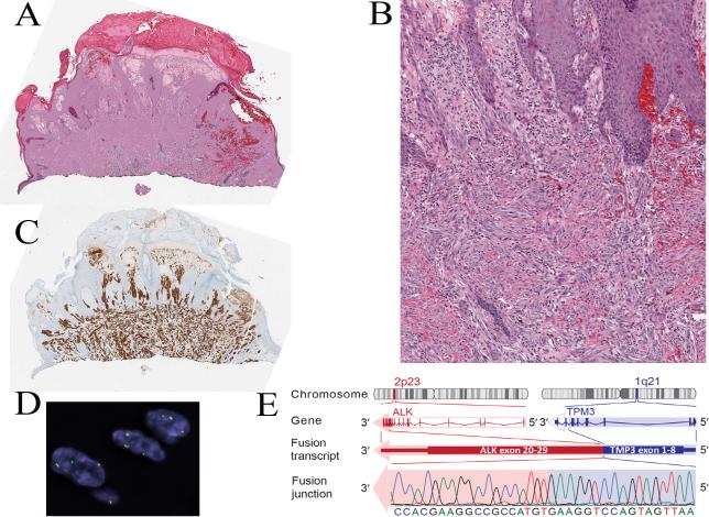
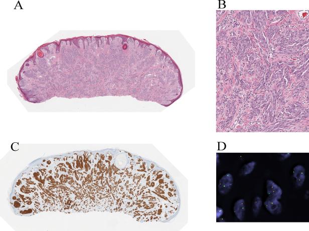
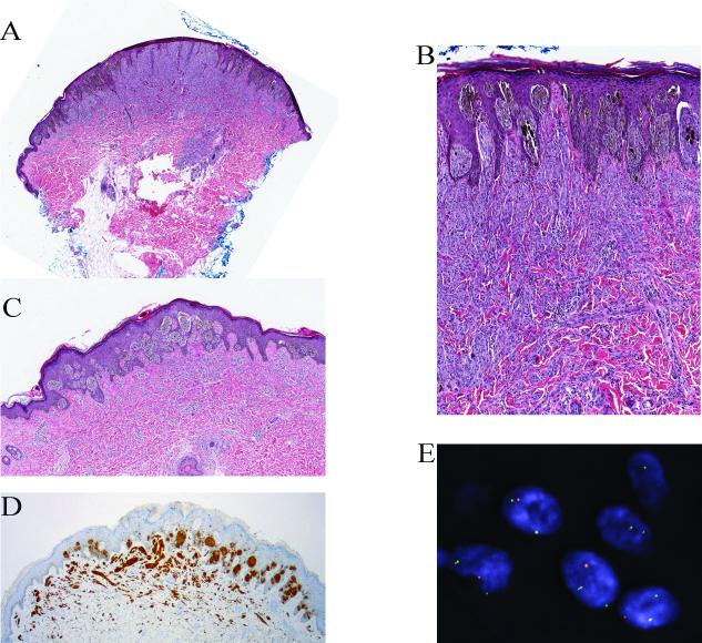
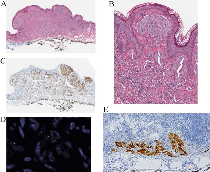
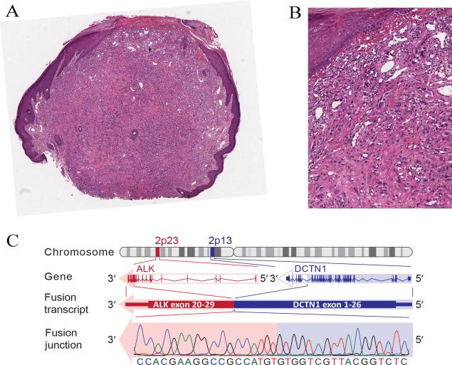
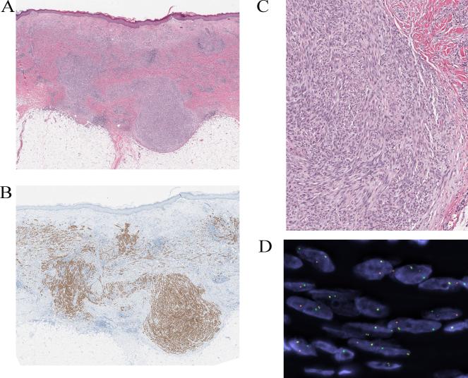
Spitz tumors represent a group of melanocytic neoplasms that typically affect young individuals. Microscopically, the lesions are composed of cytologically distinct spindle and epithelioid melanocytes, with a range in the architectural display or the cells, their nuclear features, and secondary epidermal or stromal changes. Recently, kinase fusions have been documented in a subset of Spitz tumors, but there is limited information on the clinical and pathologic features associated with those lesions. Here, we report a series of 17 patients (9 male, 8 female) with spitzoid neoplasms showing ALK fusions (5 Spitz nevi and 12 atypical Spitz tumors). The patients' ages ranged from 2 years to 35 years (mean=17 y; median=16 y). Most lesions were located on the lower extremities and presented clinically as polypoid nodules. All tumors were compound melanocytic proliferations with a predominant intradermal growth. Tumor thickness ranged from 1.1 to 6 mm (mean=2.9 mm; median=2.5 mm). The most characteristic histopathologic feature of the tumors (seen in all but 2 lesions) was a plexiform dermal growth of intersecting fascicles of fusiform melanocytes. All but 2 tumors were amelanotic. All tumors were strongly immunoreactive for ALK. The ALK rearrangements were confirmed in all cases by fluorescence in situ hybridization (FISH), and the fusion partner was determined by quantitative polymerase chain reaction as TPM3 (tropomyosin 3) in 11 cases and DCTN1 (dynactin 1) in 6 cases. None of the 8 tumors that were analyzed by FISH for copy number changes of 6p, 6q, 9p, or 11q met criteria for melanoma. Two patients underwent a sentinel lymph node biopsy, and in both cases melanocyte nests were found in the subcapsular sinus of the node. Array comparative genomic hybridization of these 2 tumors revealed no chromosomal gains or losses. In conclusion, our study revealed that Spitz nevi/tumors with ALK rearrangement show a characteristic plexiform morphology and that ALK immunohistochemistry and FISH enable the accurate identification of this morphologic and genetic distinct subset of spitzoid neoplasms.


Figures

Figure 1
Figure 1
Spitz nevus with a TPM3-ALK fusion from the chin of a 5-year-old boy (case 1). A, Silhouette of an irritated nevus with epidermal hyperplasia and hemorrhagic crust associated with focal ulceration. B, Proliferation of cytologically bland spindle cells (hematoxylin and eosin-stained section). C, The tumor cells are immunoreactive for ALK. D, Fluorescence in situ hybridization (FISH) demonstrates the ALK gene rearrangement by the individual green and orange signals using breakpoint flanking probes. E, TPM3-ALK fusion. ALK is located on chromosome 2p23. Due to genomic rearrangements, exon 1-8 of TPM3 is fused with exon 20-29 of ALK, which contains the tyrosine kinase domain.
Figure 2
Figure 2
Spitz nevus with a TPM3-ALK fusion from the leg of a 25-year-old man (case 2). A, Silhouette of an amelanotic polypoid compound Spitz nevus (hematoxylin and eosin-stained section) with evidence of maturation. B, Proliferation of cytologically bland spindle and epithelioid melanocytes. C, The tumor cells are positive for ALK. D, FISH confirms the ALK rearrangements using breakpoint flanking probes by the individual green and red signals.
Figure 3
Figure 3
Partly pigmented compound Spitz nevus with a TPM3-ALK fusion from the thigh of an 11-year-old girl (case 3). A, Wedge-shaped silhouette of a compound spindle cell melanocytic proliferation with epidermal hyperplasia (hematoxylin and eosin-stained section). B, The junctional component shows features of a pigmented spindle cell nevus. C, Deeper section of the lesion, which was adjacent to the section used for immunohistochemistry. The junctional melanocytic proliferation shows a predominant nested pattern and is pigmented. The intradermal melanocytes are amelanotic and display a plexiform growth pattern. D, The tumor cells are positive for ALK in immunohistochemistry. E, FISH confirms the ALK rearrangement.
Figure 4
Figure 4
Compound Spitz tumor with a TPM3-ALK fusion and positive SLN from the ear of a 35-year-old man (case 4). A, Silhouette of a polypoid compound melanocytic proliferation (hematoxylin and eosin-stained section). B, Amelanotic spindle and epithelioid melanocytic proliferation with evidence of maturation. C, The tumor cells are immunoreactive for ALK. D, FISH confirms the ALK rearrangement. E, Spitzoid melanocyte deposits were found in the subcapsular sinus of the sentinel lymph node (immunohistochemical stain for S100 protein).
Figure 5
Figure 5
Compound Spitz tumor with a DCTN1-ALK fusion from the arm of a 19-year-old man (case 16). A, Polypoid compound melanocytic tumor, focally ulcerated (hematoxylin and eosin-stained section). B, Proliferation of amelanotic predominantly large epithelioid melanocytes with nuclear atypia. C, DCTN1-ALK kinase fusion. ALK is located on chromosome 2p23 and DCTN1 on chromosome 2p13. Due to genomic rearrangements, exon 1-26 of DCTN1 is fused with exon 20 to 29 of ALK, which contains the tyrosine kinase domain. The in-frame junction of the fusion transcript was confirmed with Sanger sequencing.
Figure 6
Figure 6
Compound Spitz tumor with a DCTN1-ALK fusion from the thigh of a 9-year-old girl (case 13). A, Plaque-like intradermal melanocytic proliferation associated with a superficial dermal biopsy-related scar. The lesion shows a plexiform growth of amelanotic spindle cells with bulbous nodular growth into the superficial subcutis (hematoxylin and eosin-stained section). B, The bulbous nodule is composed of a dense proliferation of fusiform melanocytes. C, The melanocytes are immunoreactive for ALK. D, FISH confirms the ALK rearrangement.

Similar articles

Cited by

References

    1. Zedek DC, McCalmont TH. Spitz nevi, atypical spitzoid neoplasms, and spitzoid melanoma. Clin Lab Med. 2011;31(2):311–20. - PubMed
    1. Bastian BC, LeBoit PE, Pinkel D. Mutations and copy number increase of HRAS in Spitz nevi with distinctive histopathological features. Am J Pathol. 2000;157(3):967–72. - PMC - PubMed
    1. Bastian BC, et al. Molecular cytogenetic analysis of Spitz nevi shows clear differences to melanoma. J Invest Dermatol. 1999;113(6):1065–9. - PubMed
    1. Fullen DR, et al. BRAF and NRAS mutations in spitzoid melanocytic lesions. Mod Pathol. 2006;19(10):1324–32. - PubMed
    1. Gerami P, Busam KJ. Cytogenetic and mutational analyses of melanocytic tumors. Dermatol Clin. 2012;30(4):555–66. v. - PubMed

Publication types

MeSH terms