

Transmission dynamics of Borrelia turicatae from the arthropod vector
- PMID: 24699275
- PMCID: PMC3974661
- DOI: 10.1371/journal.pntd.0002767
Transmission dynamics of Borrelia turicatae from the arthropod vector
Abstract
Background: With the global distribution, morbidity, and mortality associated with tick and louse-borne relapsing fever spirochetes, it is important to understand the dynamics of vector colonization by the bacteria and transmission to the host. Tick-borne relapsing fever spirochetes are blood-borne pathogens transmitted through the saliva of soft ticks, yet little is known about the transmission capability of these pathogens during the relatively short bloodmeal. This study was therefore initiated to understand the transmission dynamics of the relapsing fever spirochete Borrelia turicatae from the vector Ornithodoros turicata, and the subsequent dissemination of the bacteria upon entry into murine blood.
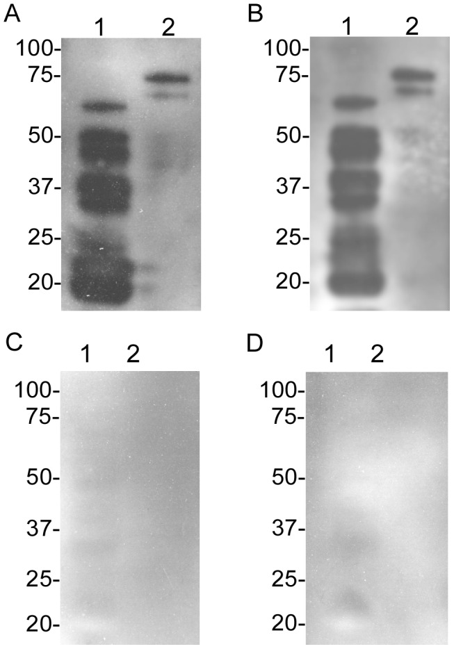
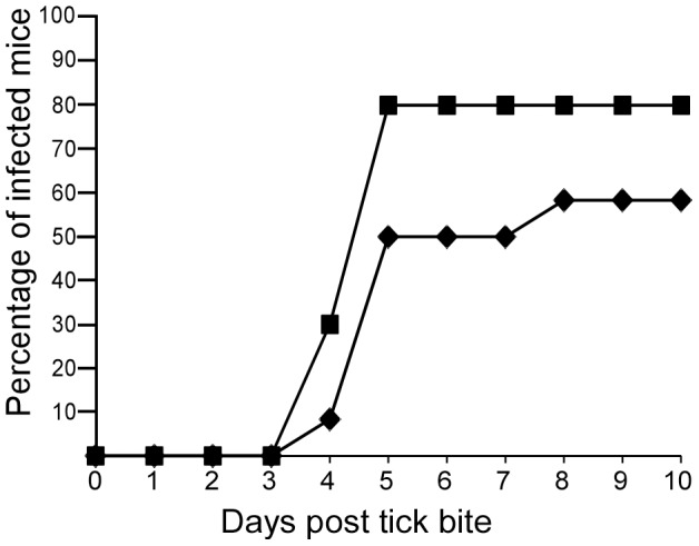
Methodology/principal findings: To determine the minimum number of ticks required to transmit spirochetes, one to three infected O. turicata were allowed to feed to repletion on individual mice. Murine infection and dissemination of the spirochetes was evaluated by dark field microscopy of blood, quantitative PCR, and immunoblotting against B. turicatae protein lysates and a recombinant antigen, the Borrelia immunogenic protein A. Transmission frequencies were also determined by interrupting the bloodmeal 15 seconds after tick attachment. Scanning electron microscopy (SEM) was performed on infected salivary glands to detect spirochetes within acini lumen and excretory ducts. Furthermore, spirochete colonization and dissemination from the bite site was investigated by feeding infected O. turicata on the ears of mice, removing the attachment site after engorment, and evaluating murine infection.
Conclusion/significance: Our findings demonstrated that three ticks provided a sufficient infectious dose to infect nearly all animals, and B. turicatae was transmitted within seconds of tick attachment. Spirochetes were also detected in acini lumen of salivary glands by SEM. Upon host entry, B. turicatae did not require colonization of the bite site to establish murine infection. These results suggest that once B. turicatae colonizes the salivary glands the spirochetes are preadapted for rapid entry into the mammal.
Conflict of interest statement
The authors have declared that no competing interest exist.
Figures


References
-
- Sonenshine DE, Mather TN (1994) Ecological Dynamics of Tick-Borne Zoonoses: Oxford University Press, Inc. 447 p.
-
- Hovius JW, van Dam AP, Fikrig E (2007) Tick-host-pathogen interactions in Lyme borreliosis. Trends Parasitol 23: 434–438. - PubMed
-
- Piesman J (1991) Experimental acquisition of the Lyme disease spirochete, Borrelia burgdorferi, by larval Ixodes dammini (Acari: Ixodidae) during partial blood meals. J Med Entomol 28: 259–262. - PubMed
-
- Piesman J (1993) Dynamics of Borrelia burgdorferi transmission by nymphal Ixodes dammini ticks. J Infect Dis 167: 1082–1085. - PubMed
-
- Piesman J (1995) Dispersal of the Lyme disease spirochete Borrelia burgdorferi to salivary glands of feeding nymphal Ixodes scapularis (Acari: Ixodidae). J Med Entomol 32: 519–521. - PubMed
Publication types
MeSH terms
Grants and funding
LinkOut - more resources
Full Text Sources
Other Literature Sources
Medical
Molecular Biology Databases
