. 2014 Apr 3:5:3546.
doi: 10.1038/ncomms4546.

Porphyrin-phospholipid liposomes permeabilized by near-infrared light

Affiliations
Free PMC article

Porphyrin-phospholipid liposomes permeabilized by near-infrared light

Kevin A Carter et al. Nat Commun. .
Free PMC article

Abstract

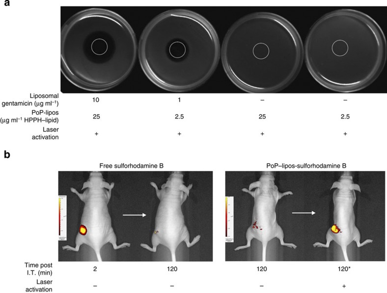
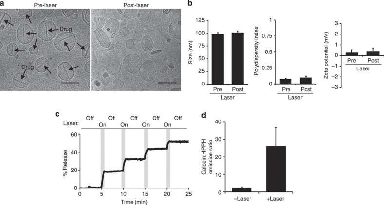
The delivery of therapeutic compounds to target tissues is a central challenge in treating disease. Externally controlled drug release systems hold potential to selectively enhance localized delivery. Here we describe liposomes doped with porphyrin-phospholipid that are permeabilized directly by near-infrared light. Molecular dynamics simulations identified a novel light-absorbing monomer esterified from clinically approved components predicted and experimentally demonstrated to give rise to a more stable porphyrin bilayer. Light-induced membrane permeabilization is enabled with liposomal inclusion of 10 molar % porphyrin-phospholipid and occurs in the absence of bulk or nanoscale heating. Liposomes reseal following laser exposure and permeability is modulated by varying porphyrin-phospholipid doping, irradiation intensity or irradiation duration. Porphyrin-phospholipid liposomes demonstrate spatial control of release of entrapped gentamicin and temporal control of release of entrapped fluorophores following intratumoral injection. Following systemic administration, laser irradiation enhances deposition of actively loaded doxorubicin in mouse xenografts, enabling an effective single-treatment antitumour therapy.


Figures

Figure 1
Figure 1. MD simulations of a stable porphyrin bilayer.
(a) Chemical structures of pyro–lipid 1 and HPPH-lipid 2. Differences are shown in bold. (b) Cross-sectional area of bilayers formed entirely from HPPH–lipid in a 500 ns, 128 monomer MD simulation. One of the HPPH monomers is depicted in wire frame format. (c) HPPH–lipid and pyro–lipid density (excluding water contribution) post 500 ns MD simulation. (d) Evolution of hydrogen bonds formed during MD simulation. Total (intermolecular plus intramolecular) and intermolecular hydrogen bonds for each type of porphyrin–lipid are indicated. (e) Chain order parameter (SZZ) for porphyrin–lipids following 500 ns MD simulation. SZZ indicates order of lipid chain with respect to bilayer normal vector. Error bars show range for last two adjacent 50 ns block means. (f) Stable cargo retention from bilayers of HPPH–lipid, but not pyro–lipid. Both monomers were synthesized and assembled into nanovesicles loaded with calcein at self-quenching concentrations. Nanovesicles were separated from unentrapped calcein before assessing fluorescence. Fluorescence emission of retained calcein in nanovesicles is shown prior or following addition of detergent (det.; 0.25% Triton X-100).
Figure 2
Figure 2. NIR-mediated liposomal cargo unloading in the absence of heating.
(a) Calcein-loaded PoP-liposomes were formed from 5% PEG–lipid, 35% cholesterol and 60% DSPC. HPPH–lipid was titrated in place of DSPC as indicated. Liposomes were irradiated for 3 min with a 120 mW 658 nm laser and release was assessed. Mean±s.d. for n=3. (b,c) Calcein release (solid black line) and solution temperature (Temp; dashed blue line) was measured for PoP-liposomes in the absence (b) or presence (c) of 150 mW laser irradiation. Temperature in the solution was measured using a thermocouple. (d) ESR of a PoP-liposome sample containing 1 mol. % 5-DSA as a spin label, recorded at 50 °C. (e) Temperature dependence of ESR spectra of 5-DSA containing PoP-liposomes. (f) Evidence for lack of nanoscale heating in irradiated PoP-liposomes. The central ESR peak-to-trough width is shown for PoP-liposomes containing 5-DSA at various temperature and before (green), during (blue) and after (red) irradiation that induces permeabilization.
Figure 3
Figure 3. Stability of PoP-liposomes.
(a) Calcein-loaded PoP-liposomes doped with 10 molar (mol.) % HPPH–lipid were incubated for 10 min in saline at the indicated temperatures with or without a 3 min laser pre-treatment. Mean±s.d. for n=3. (b) Sulforhodamine B-loaded PoP-liposomes were added to hot agarose (~60 °C) before pouring and solidification. A laser was used to mediate cargo release with high spatial control and spell ‘UB’. Note the dye is distributed equally everywhere in the agarose. (c) Calcein release in liposomes formed with 10 mol. percent HPPH–lipid or free HPPH and irradiated with light. Mean±s.d. for n=3. (d) Rapid serum redistribution of liposomes containing free HPPH, but not HPPH–lipid, as judged by fluorescence unquenching. Mean±s.d. for n=3.
Figure 4
Figure 4. Spatial and temporal control of PoP-liposome permeabilization.
(a) Spatial control of B. subtilis killing with triggered antibiotic release. Gentamicin was loaded in PoP-liposomes and embedded in hot agar along with the bacteria. The indicated spot was irradiated with a 658 nm laser (200 mW cm−2) for 10 min and the plates were photographed 24 h later. (b) Temporal control of cargo release in Panc-1 xenografts. Mice were imaged following intratumoral (I.T.) injection of 5 nmol of either free sulforhodamine B or sulforhodamine B entrapped at self-quenching concentrations in PoP-liposomes. Representative images are shown of the indicated time points and conditions. Laser activation was performed after 2 h. Representative results shown with n=3 mice per treatment group.
Figure 5
Figure 5. Tunable and on-demand release of Dox in PoP-liposomes.
(a) Gel filtration demonstrating active Dox loading in PoP-liposomes. Over 95% of the Dox was loaded in Dox–PoP-liposomes using a 10:1 lipid to drug ratio at 60 °C for 1 h. (b) Gel filtration of liposomes following laser irradiation showing effective light-triggered release. (c) Tunable drug release using PoP-liposomes. Dox–PoP-liposomes were irradiated at varying times and laser powers in media containing 10% serum. Release was assessed using fluorescence. Mean±s.d. for n=3. (d) Stability of Dox–PoP-liposomes incubated in 10% serum at 37 °C for 2 days and subsequently subjected to 4 min, 300 mW laser irradiation. Mean±s.d. for n=3. (e) In vitro cell killing using Dox–PoP-liposomes. Panc-1 cells were incubated as indicated for 24 h with 10 μg ml−1 Dox following exposure to 200 mW cm−2 irradiation for 10 min. After 24 h, the media was replaced and viability was assessed 24 h later using the XTT assay. Mean±s.d. for n=8. ***Laser+Dox–PoP-liposomes induced significant inhibition of cell viability compared with all other groups based on one-way analysis of variance with post hoc Tukey’s test (P<0.001).
Figure 6
Figure 6. Insights into light-induced transient permeabilization of PoP-liposomes.
(a) Representative cryo-TEM images of Dox–PoP-liposomes before and after irradiation. Arrows indicate the formation of Dox-sulphate crystals within the liposomes. Scale bars, 100 nm. (b) Dynamic light scattering size, polydispersity index and zeta potential before and after laser-induced release of Dox–PoP-liposomes. (c) Temporary induced permeability as demonstrated by periodic laser irradiation of calcein-loaded PoP-liposomes. (d) Empty PoP-liposomes were incubated in a 2 mM calcein solution and irradiated with 120 mW laser irradiation for 3 min. Free calcein and PoP-liposome-entrapped calcein were separated with gel filtration, and the ratio of calcein emission to HPPH emission (using separate excitation and emission settings) was measured. Mean±s.d. for n=3.
Figure 7
Figure 7. Dox–PoP-liposome antitumour phototherapy.
(a) Biodistribution of Dox± laser treatment. Nude mice bearing KB tumours were i.v. injected with Dox–PoP-liposomes (10 mg kg−1 Dox) and 15 min later, the tumour was irradiated with a 658 nm laser at 200 mW cm−2 fluence rate for 12.5 min (150 J cm−2). Mean±s.d. for n=7–8 mice per group. Comparing laser-treated mice to non-laser-treated mice with a two-tailed Student’s independent sample t-test, only the tumour-associated tissues had statistically significant differences in Dox accumulation (P<0.05). (b) Kaplan–Meier survival curve for nude mice bearing KB tumours. Mice were given a single treatment when tumours reached 4–6 mm and were killed when tumours reached 10 mm in any direction. Mice were treated with Dox–PoP-liposomes (10 mg kg−1 Dox) as above or a corresponding amount of empty PoP-liposomes. n=5–7 mice per group. Based on the log-rank test, there was a statistically significant difference between the various treatment groups; and pairwise comparisons showed that Dox–PoP-lipos+laser group lived significantly longer than each other group (P<0.05).

References

    1. Peer D. et al.. Nanocarriers as an emerging platform for cancer therapy. Nat. Nanotech. 2, 751–760 (2007). - PubMed
    1. Allen T. M. & Cullis P. R. Liposomal drug delivery systems: from concept to clinical applications. Adv. Drug Deliv. Rev. 65, 36–48 (2013). - PubMed
    1. Li S.-D. & Huang L. Pharmacokinetics and biodistribution of nanoparticles. Mol. Pharm. 5, 496–504 (2008). - PubMed
    1. Alexis F., Pridgen E., Molnar L. K. & Farokhzad O. C. Factors affecting the clearance and biodistribution of polymeric nanoparticles. Mol. Pharm. 5, 505–515 (2008). - PMC - PubMed
    1. Timko B. P., Dvir T. & Kohane D. S. Remotely triggerable drug delivery systems. Adv. Mater. 22, 4925–4943 (2010). - PubMed

Publication types

MeSH terms