Review
. 2014 Apr;20(2 Cerebrovascular Disease):335-51.
doi: 10.1212/01.CON.0000446105.67173.a8.

Evaluation and management of cerebral venous thrombosis

Review

Evaluation and management of cerebral venous thrombosis

Cheryl Bushnell et al. Continuum (Minneap Minn). 2014 Apr.

Abstract

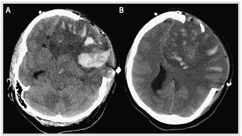
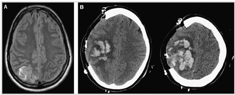
Purpose of review: Thrombosis of the dural sinus and/or cerebral veins (CVT) is a rare but potentially devastating type of stroke that tends to occur in young adults, especially women. In this article, the abbreviation CVT refers to either cerebral venous thrombosis or dural sinus thrombosis. The purpose of this review is to review the most up-to-date literature on the epidemiology, diagnosis, management, and prognosis of CVT. In addition, illustrative cases that represent the spectrum of CVT are provided.

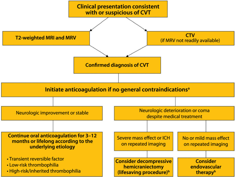
Recent findings: CVT represents about 0.5% of all strokes and can be challenging to diagnose because headache, rather than focal neurologic symptoms, is the prominent feature. The diagnosis is confirmed with MRI and magnetic resonance venogram (MRV). The mainstay of acute management is anticoagulation, although, in the cases of severe hemorrhagic conversion of a venous infarction, endovascular mechanical thrombectomy may be potentially lifesaving. The evaluation of underlying causes from transient triggers, eg, pregnancy, oral contraceptives, or infection, versus chronic triggers, eg, cancer and thrombophilia, will often influence the duration of anticoagulation. The outcomes after CVT are generally favorable, and the risk of recurrence is low.

Summary: CVT is an important diagnosis to keep in mind when evaluating patients with headache in the emergency department, and it is important that it not be overlooked because it is treatable.


Figures

Figure 5-1.
Figure 5-1.
Cerebral venous thrombosis: most commonly affected sinuses. The sagittal sinus is the most commonly affected, followed by the transverse sinuses. Modified from Saposnik G, et al, Stroke. © 2011 American Heart Association, Inc. stroke.ahajournals.org/content/42/4/1158.long.
Figure 5-2.
Figure 5-2.
A young woman presenting with headache in the emergency department. A, Plain CT shows increased attenuation involving the superior sagittal sinus, straight sinus, the confluence, and right transverse sinus (yellow arrows). B, CT venogram reveals absence of contrast in the superior sagittal sinus, straight sinus, right transverse sinus (green arrow), right sigmoid sinus, and right internal jugular vein (yellow arrow). C, Three-dimensional reconstruction of the CT venogram shows thrombosis of the superior sagittal sinus (yellow arrows).
Figure 5-3.
Figure 5-3.
A young woman with headache. A, Magnetic resonance venogram (MRV) showing an extensive thrombosis throughout the posterior two-thirds of the superior sagittal sinus, involving the torcular herophili, straight sinus, the right transverse and sigmoid sinuses, and extending inferiorly into the right internal jugular vein (arrows). The left transverse and sigmoid sinuses and left internal jugular veins are spared. B, MRV (sagittal view) showing the thrombosis of two-thirds of the superior sagittal sinus, involving the torcular herophili and the straight sinus (arrow). C, MRV at 1 year showing complete recanalization of the previously thrombosed sinuses.
Figure 5-4.
Figure 5-4.
A young pregnant woman with mastoiditis. A, Magnetic resonance venogram showing acute thrombosis of the left transverse and sigmoid sinuses. B, T1-weighted MRI of the brain showing left mastoid effusion and adjacent thrombosis that include the cortical veins of the tentorium. C, Resolution of the left transverse and sigmoid thrombosis 20 months later.
Figure 5-5.
Figure 5-5.
Algorithm for the management of cerebral venous thrombosis. CVT = cerebral venous thrombosis; MRI = magnetic resonance imaging; MRV = magnetic resonance venography; CTV = computed tomography venography; ICH = intracerebral hemorrhage. a Anticoagulation remains the principal therapy and is aimed at preventing thrombus propagation and increasing recanalization. This algorithm is not comprehensive, nor is it applicable to all clinical scenarios; patient management must be individualized. b Limited evidence is available on the benefits of decompressive hemicraniectomy and endovascular therapy for the management of cerebral venous thrombosis. Modified from Saposnik G, et al, Stroke. © 2011 American Heart Association, Inc. stroke.ahajournals.org/content/42/4/1158.long.
Figure 5-6.
Figure 5-6.
Hemorrhagic venous infarction and endovascular therapy. A, Head CT at presentation showing the large right temporal venous infarct with hemorrhagic conversion and mass effect. B, Magnetic resonance venogram (MRV) showing thrombosis of the right transverse and sigmoid sinus. C, Interventional angiogram with catheter in the right transverse sinus, status post thrombolysis and balloon angioplasty, and resolution of the thrombus.
Figure 5-7.
Figure 5-7.
Mass effect from the hemorrhagic conversion of a venous infarction (A–D, arrows) within the left frontal lobe lesion. Signs of brain herniation are evident, as is thrombosis of several left frontal cortical veins with very subtle extension in the superior sagittal sinus. The lack of visualization of residual lumen represents the occluded cortical veins and the associated severe mass effect from the adjacent brain edema (C, D, arrows).
Figure 5-8.
Figure 5-8.
Large area of altered signal and swelling with restricted diffusion (A, B) involving the cortex and underlying white matter of most of the left frontal lobe. Several areas of hypointense signal are present, more obvious on the gradient-echo and susceptibility-weighted imaging sequence in keeping with multiple hemorrhagic foci (D). The magnetic resonance venogram (C) shows few multifocal filling defects involving the superior sagittal sinus. These defects are nonobstructive. A focal severe attenuation of the central lumen of the superior sagittal sinus (B, C, thick arrows) is present, as is a paucity of expected opacified left convexity cortical veins compared with the right frontal lobe (thin arrow).
Figure 5-9.
Figure 5-9.
Noncontrast CT post craniectomy showing further enlargement of the large left hemispheric lesion and associated multifocal hemorrhagic components and brain herniating through the craniectomy site with persistent signs of severe mass effect on the midline structures and basal cisterns. A, Inferior section, and B, superior section, both of which illustrate the mass effect and the hemorrhagic conversion.
Figure 5-10.
Figure 5-10.
Seizures and intracerebral hemorrhage due to cerebral venous thrombosis in the puerperium. A, Fluid-attenuated inversion recovery (FLAIR) T2 MRI shows hyperintensity in the right posterior parietal region consistent with acute cortical vein thrombosis. B, A head CT post hemicraniectomy demonstrates a large right frontoparietal intraparenchymal hemorrhage with associated mass effect and mild herniation of the brain through the craniectomy defect.

References

    1. Bousser MG,, Ferro JM. Cerebral venous thrombosis: an update. Lancet Neurol 2007; 6 (2): 162–170. - PubMed
    1. Ferro J,, Canhao P,, Stam J, et al Prognosis of cerebral vein and dural sinus thrombosis: results of the International Study on Cerebral Vein and Dural Sinus Thrombosis (ISCVT). Stroke 2004; 35 (3): 664–670. - PubMed
    1. deVeber G,, Andrew M,, Adams C, et al Cerebral sinovenous thrombosis in children. N Engl J Med 2001; 345 (6): 417–423. - PubMed
    1. Stam J. Thrombosis of the cerebral veins and sinuses. N Engl J Med 2005; 352 (17): 1791–1798. - PubMed
    1. Crassard I,, Bousser MG. Headache in patients with cerebral venous thrombosis [in French]. Rev Neurol (Paris) 2005; 161 (6–7): 706–708. - PubMed

Publication types

MeSH terms