. 2014 Apr 3;10(4):e1004053.
doi: 10.1371/journal.ppat.1004053. eCollection 2014 Apr.

Alveolar macrophages are essential for protection from respiratory failure and associated morbidity following influenza virus infection

Affiliations

Alveolar macrophages are essential for protection from respiratory failure and associated morbidity following influenza virus infection

Christoph Schneider et al. PLoS Pathog. .

Abstract

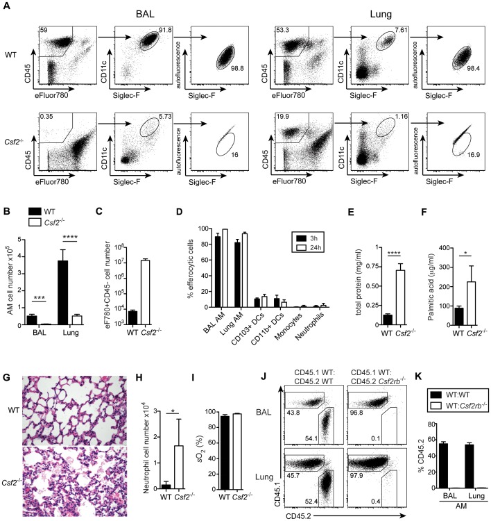
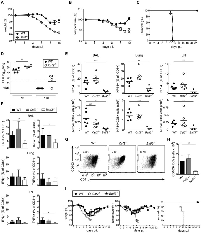
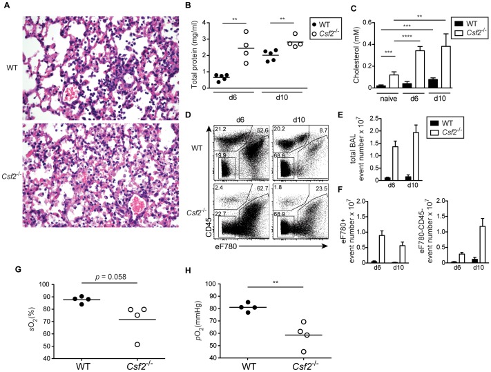
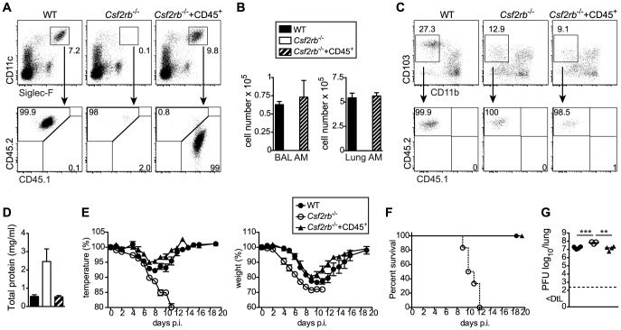
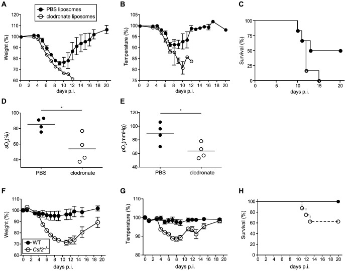
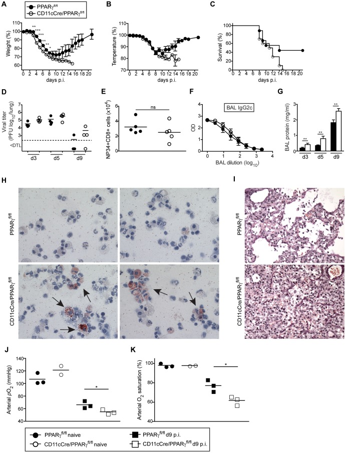
Alveolar macrophages (AM) are critical for defense against bacterial and fungal infections. However, a definitive role of AM in viral infections remains unclear. We here report that AM play a key role in survival to influenza and vaccinia virus infection by maintaining lung function and thereby protecting from asphyxiation. Absence of AM in GM-CSF-deficient (Csf2-/-) mice or selective AM depletion in wild-type mice resulted in impaired gas exchange and fatal hypoxia associated with severe morbidity to influenza virus infection, while viral clearance was affected moderately. Virus-induced morbidity was far more severe in Csf2-/- mice lacking AM, as compared to Batf3-deficient mice lacking CD8α+ and CD103+ DCs. Csf2-/- mice showed intact anti-viral CD8+ T cell responses despite slightly impaired CD103+ DC development. Importantly, selective reconstitution of AM development in Csf2rb-/- mice by neonatal transfer of wild-type AM progenitors prevented severe morbidity and mortality, demonstrating that absence of AM alone is responsible for disease severity in mice lacking GM-CSF or its receptor. In addition, CD11c-Cre/Ppargfl/fl mice with a defect in AM but normal adaptive immunity showed increased morbidity and lung failure to influenza virus. Taken together, our results suggest a superior role of AM compared to CD103+ DCs in protection from acute influenza and vaccinia virus infection-induced morbidity and mortality.


Conflict of interest statement

The authors have declared that no competing interests exist.

Figures

Figure 1
Figure 1. Cell-intrinsic requirement of GM-CSF for the development of alveolar macrophages.
(A) Identification of live (eFluor780) AM (CD45+CD11c+Siglec-F+ autofluorescenthigh) in BAL and lung of WT and Csf2 −/− mice. (B) Absolute cell numbers of AM. (C) Quantification of dead CD45eFluor780+ cells in the BAL. (D) Efferocytosis of i.t. instilled apoptotic thymocytes by indicated populations of myeloid cells. Values shown depict percentages of efferocytic cells. (E and F) Concentrations of total protein (E) and palmitic acid (F) were measured in the BAL of WT and Csf2 −/− mice. (G) Panels show H&E-stained histological lung sections of WT and Csf2 −/− mice. (H) Absolute cell numbers of neutrophils (CD11b+CD11cGr-1+) in the BAL. (I) Arterial oxygen saturation in WT and Csf2 −/− mice. (J and K) Mixed BM chimeras (1∶1 mixture of CD45.1+WT∶CD45.2+ Csf2rb −/− or CD45.1+WT∶CD45.2+WT) were analyzed for the contribution of CD45.1 and CD45.2 BM to the development of AM. (J) Dot plots show the percentage of CD45.1+WT and CD45.2+ Csf2rb −/− (or control CD45.2+WT) cells among CD11c+Siglec-F+ AM in the BAL and lung. (K) Bar graphs display the frequency of CD45.2+WT and CD45.2+ Csf2rb −/− AM as gated in (J). The mean ± SD is shown.
Figure 2
Figure 2. GM-CSF promotes development of CD103+ DCs and expression of CD103 in young and adult mice, respectively.
Analysis of lung DC subsets in (A–C) 6–8 week-old and (D–E) 6-day-old mice. (A, D) Gated cells in dot plots show CD103+ and CD11b+ DCs with values indicating percentages among CD11c+Siglec-F lung DCs. Cells were pre-gated on CD45+eFluor780 viable cells. (B,E) Bar graphs depict total number of CD103+ and CD11b+ DCs in the lung. (C) Mean fluorescence intensity (MFI) of CD103 expression on CD103+CD11b DCs. Values shown represent the mean ± SD (n = 3–4). (F) Analysis of lung DC subsets in mixed BM chimeras generated by transfer of 1∶1 mixture of CD45.1+WT∶CD45.2+ Csf2rb −/− or control CD45.1+WT∶CD45.2+WT into lethally irradiated recipients. Bar graphs show the percentage of CD45.2+WT and CD45.2+ Csf2rb −/− cells among CD103+ and CD11b+ DCs in the lung. The mean ± SD is shown (n = 3).
Figure 3
Figure 3. Csf2 −/− mice succumb to influenza virus infection despite intact antiviral T and B cell responses.
(A–I) Indicated groups of mice were infected i.t. with PR8 influenza virus (50 pfu unless otherwise specified). Loss of body weight (A) and temperature (B) was monitored. (C) Survival after infection with 100 pfu PR8. Values indicate mean ± SEM of 8–10 mice per group. (D) Virus titers in the lung at days 6 and 10 p.i. were measured by plaque-assay. (E–H) Comparison of immune responses in Csf2 −/−, Batf3 −/− and WT mice at day 10 post infection. (E) Percentages (upper panel) and total numbers (lower panel) of influenza NP34-specific CD8+ T cells in the BAL, lung and lung-draining LN. (F) Virus-specific IFNγ and TNFα production in CD8+ T cells of the BAL, lung and LN was analyzed by restimulation with virus-loaded BMDCs. (G and H) CD103+ DCs in the lung at d10 post-infection were gated on eF780CD45+CD11c+MHCII+B220Siglec-F cells. Dot plots show the frequencies of CD103+ DCs of individual mice representative for the group (G) and bar graphs display total numbers (H). Values indicate mean ± SD of 5–6 mice per group. (I) WT, Csf2 −/− and Batf3 −/− mice were monitored for body weight, body temperature and the survival during the course of infection. Data show mean ± SEM.
Figure 4
Figure 4. Defective gas exchange and respiratory failure following influenza virus infection in Csf2 −/− mice lacking AM.
WT and Csf2 −/− mice were infected i.t. with 50 pfu PR8 influenza virus. (A) Panels show H&E-stained histological lung sections of day 10-infected WT and Csf2 −/− mice. The concentration of total protein (B) and cholesterol (C) in the BAL was determined at the indicated time points after infection. (D) Percentages of dead cells (eFluor780+) and debris (eFluor780CD45) in the BAL were determined by flow cytometry at d6 and d10. Bar graphs show the total BAL event number (E) and numbers of eFluor780+ or eFluor780CD45 events (F). Shown is the mean ± SD of 4–5 mice per group. Arterial blood oxygen saturation (G) and O2 partial pressure (H) was measured in infected animals at day 9. Symbols represent values of individual mice and the mean is shown.
Figure 5
Figure 5. Selective restoration of AM development in Csf2rb −/− mice prevents severe morbidity and mortality following influenza virus infection.
Whole CD45+ cells containing progenitors of AM were sorted from lungs of CD45.1+ E18.5 embryos by flow cytometry and transferred intranasally into neonatal CD45.2+ Csf2rb −/− mice. (A) Six weeks after transfer, recipient mice were analyzed for the presence of donor-derived AM. (B) Bar graphs display the total AM cell number in the BAL and lung. (C) Dot plots depict the expression of CD103 and CD11b on CD11c+MHCII+Siglec-F lung DCs and frequencies of CD45.1+ (donor-derived) and CD45.2+ (recipient-derived) cells among CD103+ DCs are shown. (D–G) Eight weeks after transfer, recipient mice were infected with 50 pfu PR8 influenza virus. (D) Total protein concentration in the BAL at d5 after infection. (E) Loss of body weight and temperature and (F) survival was monitored during the course of infection (mean ± SEM of 6–9 mice per group). (G) Lung virus titer at day 5 p.i.
Figure 6
Figure 6. Increased morbidity and respiratory failure in mice upon selective depletion of alveolar macrophages prior to pulmonary virus infection.
(A–E) Mice were treated with clodronate or control PBS liposomes 2 days prior to infection with 50 pfu PR8 influenza virus. Loss of body weight (A) and temperature (B) was monitored. (C) Survival was assessed over a period of 3 weeks after infection. Values indicate mean ± SEM of 6 mice per group. Arterial blood oxygen saturation (D) and O2 partial pressure (E) was measured in infected animals at d8. (F–H) WT and Csf2 −/− mice were infected with 104 pfu vaccinia virus WR i.t. Loss of body weight (F), loss of temperature (G) and survival (H) was monitored. Values indicate mean ± SEM of 7–8 mice per group.
Figure 7
Figure 7. CD11c-Cre/Ppargfl/fl mice have a reduced resistance to influenza virus infection despite an intact antiviral adaptive response.
Pparg fl/fl and CD11c-Cre/Pparg fl/fl mice were infected i.t. with 250 pfu PR8 influenza virus. Loss of body weight (A), temperature (B) and survival (C) was monitored. Values indicate mean ± SEM of 9–10 mice per group. (D–K) For the characterization of the anti-viral immune response, Pparg fl/fl and CD11cCre/Pparg fl/fl mice were infected i.t. with 50 pfu PR8 influenza virus. Symbols represent values of individual mice and the mean is indicated. (D) The virus titers in the lung were determined at d3, d5 and d9 after infection. (E) Total numbers of influenza NP34-specific CD8+ T cells in the BAL were analyzed at d10 post-infection. (F) Influenza HA-specific IgG2c antibody concentrations in the BAL at d13 post-infection were determined by ELISA. Values indicate mean ± SEM of 4–5 mice per group. (G) Total protein in the BAL was measured at indicated time points. (H) Cytospins of BAL isolated from infected mice at d9 were stained with Oil Red O. Micrographs were taken at 63× magnification. Representative pictures of 2 individual mice are shown. (I) Panels show lung sections of day 9-infected mice stained using the Verhoeff-Van Gieson protocol. (J and K) Lung function was measured in naïve and day 9-infected animals. Arterial blood oxygen partial pressure (J) and O2 saturation (K) are shown.
Figure 8
Figure 8. Influenza infection potently induces expression of interferon-regulated antiviral factors in AM.
(A) Mice were infected with 50 pfu PR8 influenza virus. Intracellular NP expression was measured by flow cytometry in CD11c+autofluorescent AM isolated from BAL and lung 5 days after infection. (B) Mice were infected with 106 pfu NS1-GFP virus or 103 pfu PR8. GFP expression was analyzed in AM isolated from BAL and lung 5 days after infection. (C) Microarray analysis of sorted AM from lungs of naive or influenza-infected animals at d5 post-infection with 50 pfu PR8. Bar graphs show relative expression levels of various interferon-induced genes plotted as log2-fold change in AM from infected lungs compared to naïve. The mean of two microarray samples per condition is shown. For each sample, AM from two individual mice were pooled. Differences in expression levels were validated by qPCR for most of the depicted genes (i.e. Ifitm3, Ifitm6, Ifit2, Ifit3 and Ifi205).

References

    1. Trapnell BC, Whitsett JA (2002) Gm-CSF regulates pulmonary surfactant homeostasis and alveolar macrophage-mediated innate host defense. Annual review of physiology 64: 775–802. - PubMed
    1. Trapnell BC, Whitsett JA, Nakata K (2003) Pulmonary alveolar proteinosis. The New England journal of medicine 349: 2527–2539. - PubMed
    1. Seymour JF, Presneill JJ (2002) Pulmonary alveolar proteinosis: progress in the first 44 years. American Journal of Respiratory and Critical Care Medicine 166: 215–235. - PubMed
    1. Kitamura T, Tanaka N, Watanabe J, Uchida, Kanegasaki S, et al. (1999) Idiopathic pulmonary alveolar proteinosis as an autoimmune disease with neutralizing antibody against granulocyte/macrophage colony-stimulating factor. The Journal of experimental medicine 190: 875–880. - PMC - PubMed
    1. Suzuki T, Sakagami T, Rubin BK, Nogee LM, Wood RE, et al. (2008) Familial pulmonary alveolar proteinosis caused by mutations in CSF2RA. The Journal of experimental medicine 205: 2703–2710. - PMC - PubMed

Publication types

MeSH terms

Substances