. 2014 Apr 3;10(4):e1004061.
doi: 10.1371/journal.ppat.1004061. eCollection 2014 Apr.

The pathogenic mechanism of the Mycobacterium ulcerans virulence factor, mycolactone, depends on blockade of protein translocation into the ER

Affiliations

The pathogenic mechanism of the Mycobacterium ulcerans virulence factor, mycolactone, depends on blockade of protein translocation into the ER

Belinda S Hall et al. PLoS Pathog. .

Abstract

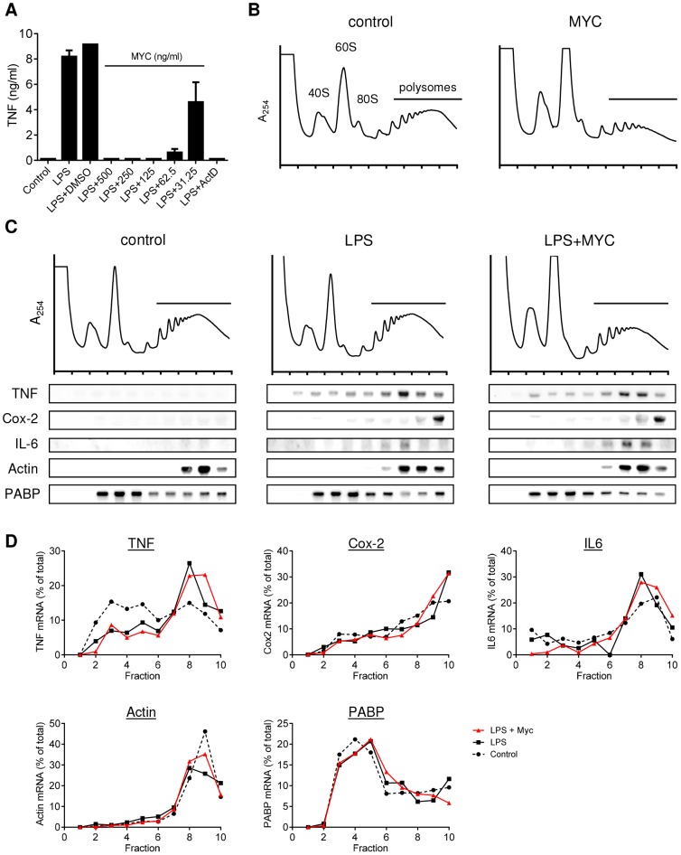
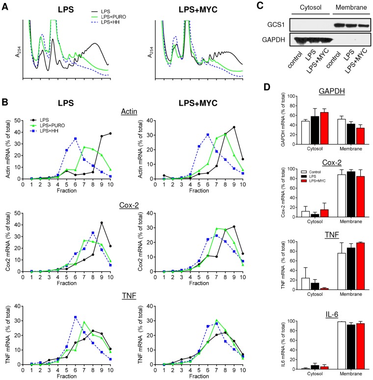
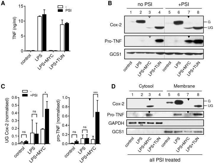
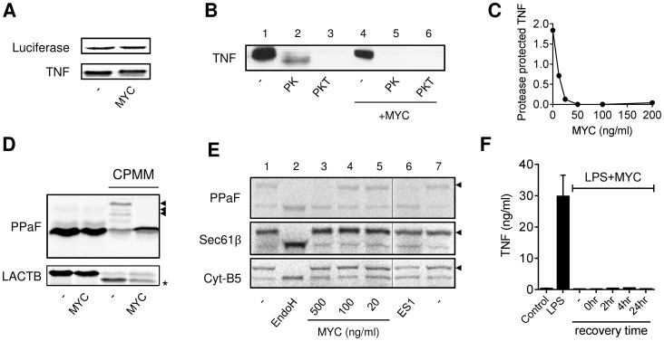
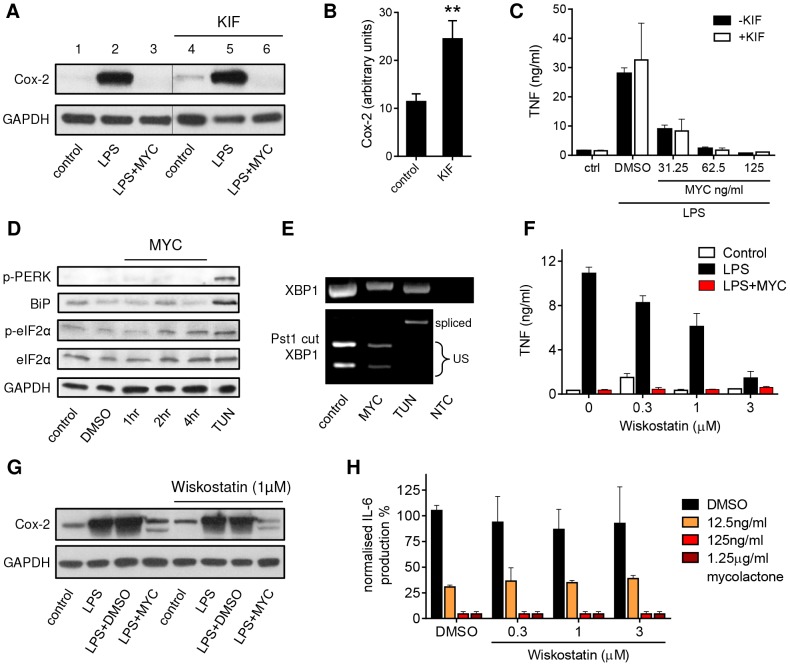
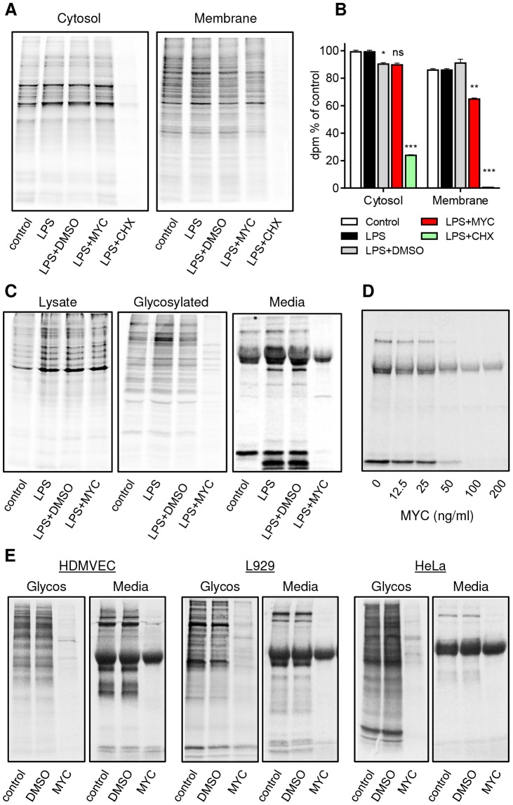
Infection with Mycobacterium ulcerans is characterised by tissue necrosis and immunosuppression due to mycolactone, the necessary and sufficient virulence factor for Buruli ulcer disease pathology. Many of its effects are known to involve down-regulation of specific proteins implicated in important cellular processes, such as immune responses and cell adhesion. We have previously shown mycolactone completely blocks the production of LPS-dependent proinflammatory mediators post-transcriptionally. Using polysome profiling we now demonstrate conclusively that mycolactone does not prevent translation of TNF, IL-6 and Cox-2 mRNAs in macrophages. Instead, it inhibits the production of these, along with nearly all other (induced and constitutive) proteins that transit through the ER. This is due to a blockade of protein translocation and subsequent degradation of aberrantly located protein. Several lines of evidence support this transformative explanation of mycolactone function. First, cellular TNF and Cox-2 can be once more detected if the action of the 26S proteasome is inhibited concurrently. Second, restored protein is found in the cytosol, indicating an inability to translocate. Third, in vitro translation assays show mycolactone prevents the translocation of TNF and other proteins into the ER. This is specific as the insertion of tail-anchored proteins into the ER is unaffected showing that the ER remains structurally intact. Fourth, metabolic labelling reveals a near-complete loss of glycosylated and secreted proteins from treated cells, whereas cytosolic proteins are unaffected. Notably, the profound lack of glycosylated and secreted protein production is apparent in a range of different disease-relevant cell types. These studies provide a new mechanism underlying mycolactone's observed pathological activities both in vitro and in vivo. Mycolactone-dependent inhibition of protein translocation into the ER not only explains the deficit of innate cytokines, but also the loss of membrane receptors, adhesion molecules and T-cell cytokines that drive the aetiology of Buruli ulcer.


Conflict of interest statement

The authors have declared that no competing interests exist.

Figures

Figure 1
Figure 1. Mycolactone does not change the polysomal association of proinflammatory mRNAs.
A. RAW264.7 cells were incubated for 1+/−various concentrations of mycolactone (MYC as indicated), 0.5 µg/ml Actinomycin D (Act D) or 0.0125% DMSO then stimulated or not with LPS for 4 hr. Supernatant TNF levels were measured by ELISA (mean±SEM of triplicate assays). B–D. RAW264.7 cells were incubated for 1 hr+/−125 ng/ml mycolactone (MYC) then stimulated or not with LPS. After 4 hrs cells were harvested and lysed in the presence of CHX. B and C. Polysomes were separated on a 10–50% sucrose gradient and the profiles measured by absorbance at 254 nm. Note the increase in the 60S peak and reduced height of the polysome peaks in MYC and LPS+MYC samples. RNA was purified from gradient fractions and transcripts were detected by Northern blotting using full coding region cDNA probes for the genes indicated. D. Signal intensity was quantified by ImageJ analysis of non-saturated phosphorscreen images. Values are presented as percentage of total signal for control (dashed line), LPS (solid black line) and LPS+MYC (red line) cells. All are representative of 3 independent experiments. PABP; poly A-tract binding protein.
Figure 2
Figure 2. Proinflammatory mRNAs are actively translating in the presence of mycolactone.
RAW264.7 cells were incubated for 1+/−125 ng/ml mycolactone (MYC), then stimulated with LPS. A and B. After 4 hr 100 µg/ml puromycin (PURO) or 5 µM homoharringtonine (HH) were added for 3 min, then CHX was added before lysis and separation of polysomes on a 10–20% sucrose gradient. LPS (solid black line), LPS+PURO (solid green line) and LPS+HH (dotted blue line). A. RNA profiles measured by absorbance at 254 nm. B. Quantitation of specific mRNAs purified from each fraction and analysed by Northern blotting. Signal intensity was quantified by ImageJ analysis of non-saturated phosphorscreen images. Values are presented as percentage of total signal. C and D. Cytosolic and digitonin-resistant membrane fractions were prepared from treated cells as described. C. Western blot of cell fractions (0.5×10⁵ cell equivalents/lane). GCS1; glucosidase I (∼92 kDa), GAPDH (∼40 kDa). D. total RNA was used as a template in qRT-PCR absolute quantitation assays and is presented as % total RNA for each gene (mean±SEM). All data representative of 3 independent experiments.
Figure 3
Figure 3. Mycolactone causes degradation of TNF and Cox-2 by the 26S proteasome in the cytosol.
RAW264.7 cells were incubated +/−125 ng/ml mycolactone (MYC) or 5 µg/ml tunicamycin (TUN) for 1 hr and stimulated with LPS for 4 hrs. In certain samples 5 µM PSI was added 2 hrs after the LPS stimulation. This allowed time for the proteasome-dependent activation of NFκB required for transcriptional activation to occur before proteasome activity was inhibited (see text). A. Supernatant TNF levels from cells treated as above, measured by ELISA (mean±SEM). B. Western blot of cell lysates (0.5×10⁶ cell equivalents/lane), the dotted line separates samples with and without PSI treatment for ease of interpretation. C. Signal intensity was quantified by ImageJ analysis of non-saturated blots and normalised to LPS+TUN (not shown). Values are mean intensity for each lane±SEM of 3 independent experiments. For Cox-2 this is the unglycosylated mol wt band only (UG Cox-2). *, P<0.05; ***, P<0.001. D. Western blot of cytosolic and digitonin-resistant membrane fractions from cells treated with PSI as described above. All data representative of (or pooled from, C) 3 independent experiments. G; glycosylated Cox-2 (∼80 kDa), UG; unglycosylated Cox-2 (∼69 kDa), pro-TNF (∼26 kDa), GAPDH (∼40 KDa), GCS1; glucosidase I (∼92 KDa). Open arrowheads indicate cells treated with mycolactone but not PSI. Closed arrowheads indicate cells treated with mycolactone and PSI.
Figure 4
Figure 4. Mycolactone inhibits co-translational translocation of proteins into the ER via a mechanism that does not disrupt the structural integrity of the ER.
A–E. In vitro translation (IVT) reactions of different capped transcripts were performed as described +/− mycolactone (MYC; 200 ng/ml unless indicated otherwise), Eeyarestatin 1 (ES1; 250 µM) or DMSO (-; 0.02%). Data representative of 3 independent experiments. A. IVT of luciferase and TNF mRNAs detected by 35S incorporation and Western blot (∼26 kDa) respectively. B. IVT of TNF mRNA was performed in the presence of semi-permeabilised RAW264.7 cells then incubated with no addition (-), or Proteinase K (PK) +/−0.1% Triton-x-100 (PKT) for 1 hr at 4°C before stopping the reaction. C. Dose dependence of loss of the PK protected band. Signal intensity was quantified by ImageJ analysis of non-saturated blots and the protected band (PK) was normalised to total pro-TNF (-). D. IVT of prepro-α Factor (PPAF) and β-lactamase (LACTB) mRNAs in the absence or presence of canine pancreatic microsomal membranes (CPMM). Black arrowhead, glycosylated forms of α Factor; *, signal peptide-cleaved LACTB. E. IVT of PPAF, Sec61β and cytochrome B5 (Cyt-B5) mRNAs in the presence CPMM. After labelling with 35S as described, membranes were isolated by centrifugation through a sucrose cushion . Endoglycosidase H (EndoH) is used to confirm glycosylation of proteins (black arrowhead), the thin line represents where an empty lane was removed from the image for ease of interpretation. F. RAW264.7 cells were incubated +/−125 ng/ml mycolactone (MYC), washed and incubated without mycolactone for various periods (recovery time) before stimulating with LPS for 4 hrs. Supernatant TNF levels were measured by ELISA (mean±SEM of triplicate assays).
Figure 5
Figure 5. Mycolactone does not mediate these effects via ERAD, ER stress or WASP-activation-dependent mechanisms.
A–C. RAW264.7 cells were incubated +/−125 ng/ml mycolactone (MYC), +/−50 µM Kifunensine (KIF) or 0.0125% DMSO for 1 hr and stimulated or not with LPS for 4 hrs as indicated. A. Western blot of cell lysates (0.5×10⁶ cell equivalents/lane); the thin line represents where an empty lane was removed from the image for ease of interpretation. B. Quantitation of constitutive expression of Cox-2 in lanes 1 and 4 (control and KIF respectively) Pixel intensity was determined using ImageJ software and normalised according to GAPDH. Results represent the mean±SEM (n = 3). C. Supernatant TNF levels measured by ELISA (mean±SEM). D. RAW264.7 cells were incubated +/−125 ng/ml mycolactone (MYC) for various periods or 5 µg/ml tunicamycin (TUN) or 0.0125% DMSO for 4 hrs. Western blot of cell lysates (0.5×10⁶ cell equivalents/lane). E. Total RNA of treated/unstimulated cells was used as a template for RT-PCR of XBP-1 (upper panel) which was then digested with Pst1 and separated on a 1% agarose gel (lower panel). NTC; no template control, US; unspliced. F–G. RAW264.7 cells were incubated +/−125 ng/ml mycolactone (MYC), −/+ wiskostatin at a range of concentrations or 0.0125% DMSO for 1 hr and stimulated or not with LPS for 4 hrs. F. Supernatant TNF levels measured by ELISA (mean±SEM). G. Western blot of cell lysates (0.5×10⁶ cell equivalents/lane). H. HeLa cells were incubated with a range of doses of mycolactone (MYC) and wiskostatin or 0.0125% DMSO for 1 hr and stimulated or not with 10 ng/ml IL-1β. IL-6 in supernatants was measured by ELISA (mean±SEM of production, normalised to IL-6 production in otherwise untreated, IL1β stimulated cells). Cox-2 (∼90 kDa), GAPDH (∼40 kDa), p-PERK (140 kDa), BiP (∼78 kDa), (p-)eIF2α (∼38 kDa).
Figure 6
Figure 6. Mycolactone specifically targets membrane and secreted proteins.
Cells in Met/Cys free medium were incubated +/−125 ng/ml mycolactone (MYC), 10 µg/ml cycloheximide (CHX) or 0.0125% DMSO for 1 hr and stimulated with LPS for 1 hr before replacement with fresh medium containing Tran35slabel for 2 hrs. Data representative of 3 independent experiments (A–D). A. Cytosolic and digitonin-resistant membrane fractions from treated RAW264.7 cells (105 cell equivalents/lane). B. Quantification of 35S incorporation in (A) by scintillation counting (mean±SEM, n = 3). *, P<0.05; **, P<0.01; ***, P<0.001. C. Total cell lysates, Concanavalin A (ConA) agarose precipitated proteins (Glycosylated) and supernatants (Media) from RAW264.7 cells. D. Dose dependence of the suppression of supernatant protein production in RAW264.7 cells. E. ConA precipitated (Glycos) and supernatant proteins (Media) from 35S labelled Human microvascular dermal epithelial cells (HDMVEC), L929 fibroblasts and Hela cells. In each case total cell labelling was comparable between samples (not shown).

References

    1. George KM, Chatterjee D, Gunawardana G, Welty D, Hayman J, et al. (1999) Mycolactone: a polyketide toxin from Mycobacterium ulcerans required for virulence. Science 283: 854–857. - PubMed
    1. Demangel C, Stinear TP, Cole ST (2009) Buruli ulcer: reductive evolution enhances pathogenicity of Mycobacterium ulcerans. Nat Rev Microbiol 7: 50–60. - PubMed
    1. Stinear TP, Mve-Obiang A, Small PL, Frigui W, Pryor MJ, et al. (2004) Giant plasmid-encoded polyketide synthases produce the macrolide toxin of Mycobacterium ulcerans. Proc Natl Acad Sci U S A 101: 1345–1349. - PMC - PubMed
    1. Hall BS, Simmonds RE (2013) Pleiotropic molecular effects of the Mycobacterium ulcerans virulence factor mycolactone underlying the cell death and immunosuppression seen in Buruli ulcer. Biochem Soc Trans 42: 177–183. - PubMed
    1. Simmonds RE, Lali FV, Smallie T, Small PL, Foxwell BM (2009) Mycolactone inhibits monocyte cytokine production by a posttranscriptional mechanism. J Immunol 182: 2194–2202. - PubMed

Publication types

MeSH terms