. 2014 Apr 15;28(8):875-87.
doi: 10.1101/gad.239194.114. Epub 2014 Apr 3.

ATR checkpoint kinase and CRL1βTRCP collaborate to degrade ASF1a and thus repress genes overlapping with clusters of stalled replication forks

Affiliations

ATR checkpoint kinase and CRL1βTRCP collaborate to degrade ASF1a and thus repress genes overlapping with clusters of stalled replication forks

Jun-Sub Im et al. Genes Dev. .

Abstract

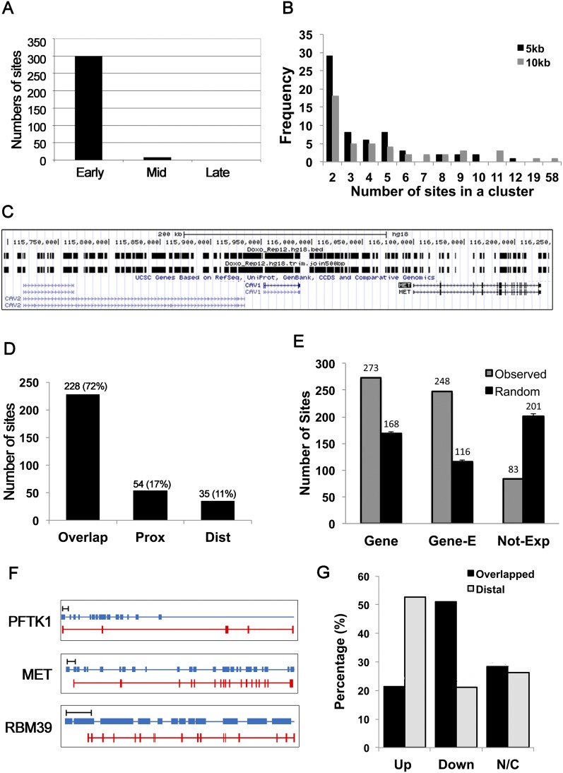
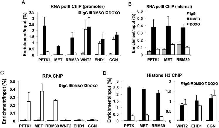
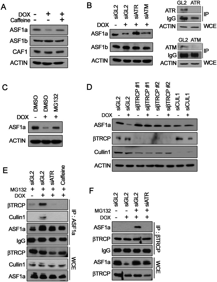
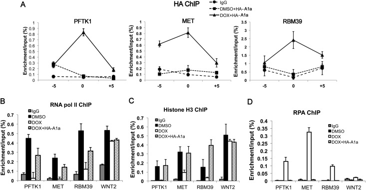
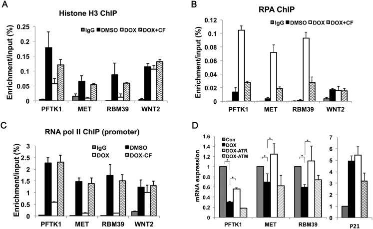
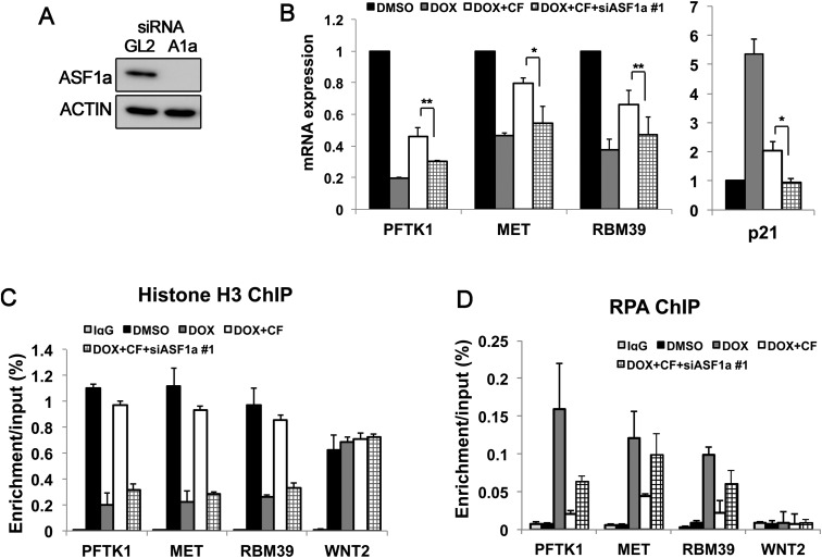
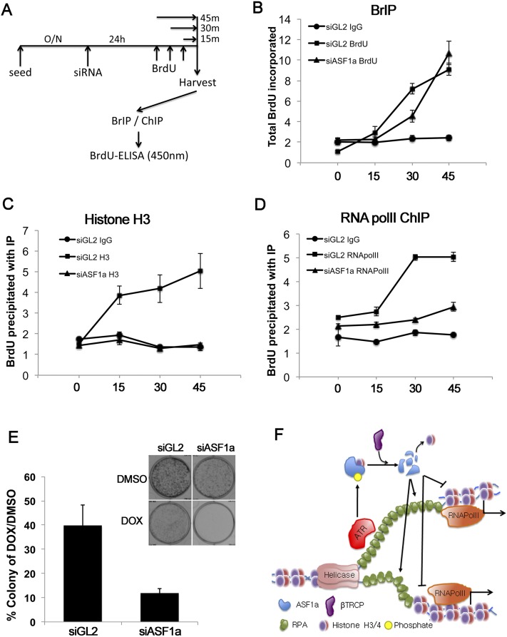
Many agents used for chemotherapy, such as doxorubicin, interfere with DNA replication, but the effect of this interference on transcription is largely unknown. Here we show that doxorubicin induces the firing of dense clusters of neoreplication origins that lead to clusters of stalled replication forks in gene-rich parts of the genome, particularly on expressed genes. Genes that overlap with these clusters of stalled forks are actively dechromatinized, unwound, and repressed by an ATR-dependent checkpoint pathway. The ATR checkpoint pathway causes a histone chaperone normally associated with the replication fork, ASF1a, to degrade through a CRL1(βTRCP)-dependent ubiquitination/proteasome pathway, leading to the localized dechromatinization and gene repression. Therefore, a globally active checkpoint pathway interacts with local clusters of stalled forks to specifically repress genes in the vicinity of the stalled forks, providing a new mechanism of action of chemotherapy drugs like doxorubicin. Finally, ASF1a-depleted cancer cells are more sensitive to doxorubicin, suggesting that the 7%-10% of prostate adenocarcinomas and adenoid cystic carcinomas reported to have homozygous deletion or significant underexpression of ASF1a should be tested for high sensitivity to doxorubicin.

Keywords: ASF1; ATR; CRL1βTRCP; S-phase checkpoint; doxorubicin; stalled forks; transcription repression.


Figures

Figure 1.
Figure 1.
DOX induced firing of clusters of dormant replication origins and decreased the mRNA expression of overlapping genes. (A) Segregation of the DOX-BrIP sites (the joined sites in Supplemental Table S1A) by whether that region replicates early, mid, or late in a normal S phase. (B) Clusters defined as BrdU-labeled tracks within 5 kb or 10 kb of each other. Sixty-two or 46 clusters were seen, respectively. The plot shows the distribution of the number of initiation sites in a cluster. (C) Representative neoreplication origin clusters on a portion of chromosome 7. Vertical bars in the top row represent individual BrIP sites. The second row shows a track where BrIP sites within 500 bp of each other were joined. This figure was captured from the University of California at Santa Cruz genome browser (http://genome.ucsc.edu; Kent et al. 2002). (D) Genes overlapping with, proximal to (within 5 kb of the TSS or TES) or distal to (>5 kb away from the TSS and TES) DOX-induced BrIP sites. The bar graph shows the number and the percentage of genes in each class. (E) The overlap of DOX-induced BrIP sites, with the indicated parts of the genome compared with a random model, reveals a significant enrichment at genes (Z = 8.1) and “expressed” genes (Gene-E; Z = 12.2) and depletion (Z = −8.3) at “not expressed” genes (Not-Exp). The Z-score represents the observed random expected/standard deviation and is explained in the Supplemental Material. (F) Representative genes that overlap with neoreplication origin clusters. The bar at the top of the figures represents 10 kb. The middle blue bar and bottom red bar represent BrdU-labeled tracks and exons of the gene, respectively. These three genes will be studied in this study as examples of genes that overlap with clusters of stalled replication forks. (G) Compared with the DMSO-treated control, genes that were induced ≥1.3-fold in DOX were considered as up-regulated (Up), whereas genes ≤0.7 are considered down-regulated (Down). Genes expressing at 0.7∼1.3 times the level in DMSO were regarded as not changed (N/C).
Figure 2.
Figure 2.
DOX produces excess RPA-bound, dechromatinized DNA near sites of stalled forks and disrupts the local recruitment of RNA polymerase II. (A,B) ChIP assay for RNA polymerase II (polII) at the promoter (A) and gene body (B) of three representative genes —(PFTK1, MET, and RBM39) overlapping with BrdU-labeled tracks and negative control genes (in A only). Each value represents a relative DNA concentration based on the standard curve of the input samples. (IgG) Normal IgG ChIP; (DMSO) anti-RNA polymerase II ChIP from DMSO-treated cells; (DOX) anti-RNA polymerase II ChIP from DOX-treated cells. Mean ± standard deviation (SD) of three measurements. (C,D) ChIP assay for RPA70 (C) and histone H3 (D) was conducted as for RNA polymerase II in A.
Figure 3.
Figure 3.
DOX induces ATR-CRL1βTRCP-dependent ASF1a degradation. (A) Immunoblots of cell lysates. ASF1a, but not ASF1b or CAF1, was decreased by DOX for 20 h, and this decrease was relieved by 5 mM caffeine added to cells from −4 h relative to DOX until the time of harvest. ACTIN was used as a loading control for the immunoblots. (B, left panel) ASF1a/b examined in cells transfected with siGL2, siATR, or siATM for 24 h and then treated with DOX for 20 h under continued exposure to siRNAs. (Right panel) Knockdown of endogenous ATR or ATM by siRNA was confirmed by immunoprecipitation (IP) and Western blot (WB). The IgG amount in each lane served as the loading control for the immunoprecipitates, and ACTIN shows equal amounts of whole -cell extracts (WCEs). (C) Cells treated with DOX for 20 h were exposed to 10 μM MG132 for 4 h before harvest. Endogenous ASF1a and ACTIN were immunoblotted. (D) Cells transfected with two different siRNAs to βTRCP for 24 h and a siRNA to Cullin1 for 48 h before DOX treatment for 20 h with continued incubation with siRNA and subjected to Western blotting for the indicated proteins and ACTIN (loading control). (E) After transfection of siGL2 or siATR (24 h) or exposure to 5 mM caffeine (4 h), cells were treated with DOX or DMSO for 20 h with continued incubation with siRNA or caffeine. Cells were harvested after exposure to 10 μM MG132 for 4 h to stabilize the ASF1a. Immunoprecipitates (top four panels) or input whole-cell extracts (bottom three panels) were immunoblotted for the indicated proteins. IgG served as a loading control for immunoprecipitates, and Cullin1 served as a loading control for whole-cell extracts. (F) An experiment similar to E, except immunoprecipitation done with anti-βTRCP antibody.
Figure 4.
Figure 4.
Ectopic ASF1a localizes near stalled forks, restoring RNA polymerase II and histone H3 while preventing RPA accumulation. (A) ChIP assay for 3HA-ASF1a at PFTK1, MET, and RBM39 was conducted after transfection with 3HA-ASF1a in the presence or absence of DOX. (−5) −5 kb from TSS; (0) TSS; (+5) +5 kb from TSS. The Y-axis indicates the amount of DNA in precipitate relative to input DNA. (B–D) RNA polymerase II (pol II) (B), histone H3 (C), or RPA (D) ChIP assay at the promoters of three genes overlapping with stalled forks and a control distal gene, WNT2.
Figure 5.
Figure 5.
The ATR-dependent checkpoint pathway is required for the transcriptional repression of genes overlapping with clusters of stalled forks. (A,B) Histone H3 (A) and RPA (B) ChIP assay for three genes overlapping with stalled forks and a control distal gene, WNT2. The rest are as in Figure 2. (DOX-CF) DOX-treated cells exposed to caffeine for 24 h before harvesting. (C) RNA polymerase II (pol II) ChIP assay at the promoters of three genes overlapping with stalled forks and a control distal gene, WNT2. (DOX-CF) DOX-treated cells exposed to caffeine for 24 h before harvesting. (D) Cells depleted of ATR or ATM by siRNA were treated with 1.5 μM DOX or DMSO (Con). The relative mRNA expression of three overlapped genes and a control gene, p21, was analyzed by qRT–PCR as in the Materials and Methods. Student’s t-test analysis, (*) P < 0.05.
Figure 6.
Figure 6.
The rescue of transcription upon inhibition of checkpoint kinase requires the restoration of ASF1a. (A) Knockdown of ASF1a by siRNA #1 for 24 h evaluated by immunoblotting. (B) mRNA levels of three genes that overlap with clusters of stalled forks analyzed by qRT–PCR. mRNA level normalized to GAPDH. Knockdown of ASF1a prevents the restoration of mRNA levels when the checkpoint is blunted with caffeine. (**) P < 0.01; (*) P < 0.05. (C,D) Chip assay for histone H3 (C) and RPA70 (D) was conducted after treatment of DOX and caffeine, as indicated, with or without knockdown of endogenous ASF1a. WNT2 was a negative control.
Figure 7.
Figure 7.
Depletion of ASF1a inhibits the loading of RNA polymerase II at newly synthesized DNA. (A–D) After 24 h of depletion of ASF1a, BrdU was added during the indicated time (shown in A). BrdU-incorporated DNA was captured by BrIP (B), histone H3 ChIP (C), or RNA polymerase II (polII) ChIP (D). BrdU-incorporated DNA eluted from BrIP or ChIP was analyzed by ELISA with anti-BrdU antibody. (E) The percentage of viable colonies after exposure of HeLa cells to DOX relative to DMSO with or without depletion of ASF1a (left) and representative examples of plates with colonies (top right). Mean ± SD from three independent experiments. (F) Schematic model of transcriptional repression of genes near DOX-induced clusters of stalled replication forks. ASF1a promotes nucleosome loading on the newly replicated DNA. This chromatinization promotes RNA polymerase II loading and suppresses unwinding or nuclease activity on the newly replicated dsDNA. ASF1a is itself degraded by ATR and CRL1βTRCP-dependent pathways activated by RPA-coated ssDNA that is present at stalled forks and that increases as more ssDNA is generated after dechromatinization of the newly replicated DNA.

References

    1. Ball HL, Myers JS, Cortez D 2005. ATRIP binding to replication protein A–single-stranded DNA promotes ATR-ATRIP localization but is dispensable for Chk1 phosphorylation. Mol Biol Cell 16: 2372–2381 - PMC - PubMed
    1. Barr SM, Leung CG, Chang EE, Cimprich KA 2003. ATR kinase activity regulates the intranuclear translocation of ATR and RPA following ionizing radiation. Curr Biol 13: 1047–1051 - PubMed
    1. Battu A, Ray A, Wani AA 2011. ASF1A and ATM regulate H3K56-mediated cell-cycle checkpoint recovery in response to UV irradiation. Nucleic Acids Res 39: 7931–7945 - PMC - PubMed
    1. Bell SP, Dutta A 2002. DNA replication in eukaryotic cells. Annu Rev Biochem 71: 333–374 - PubMed
    1. Bodley A, Liu LF, Israel M, Seshadri R, Koseki Y, Giuliani FC, Kirschenbaum S, Silber R, Potmesil M 1989. DNA topoisomerase II-mediated interaction of doxorubicin and daunorubicin congeners with DNA. Cancer Res 49: 5969–5978 - PubMed

Publication types

MeSH terms

LinkOut - more resources