

Small-angle x-ray scattering screening complements conventional biophysical analysis: comparative structural and biophysical analysis of monoclonal antibodies IgG1, IgG2, and IgG4
- PMID: 24700358
- PMCID: PMC4298811
- DOI: 10.1002/jps.23964
Small-angle x-ray scattering screening complements conventional biophysical analysis: comparative structural and biophysical analysis of monoclonal antibodies IgG1, IgG2, and IgG4
Abstract
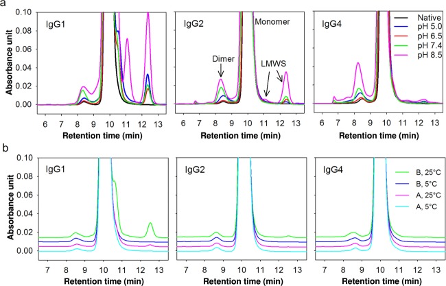
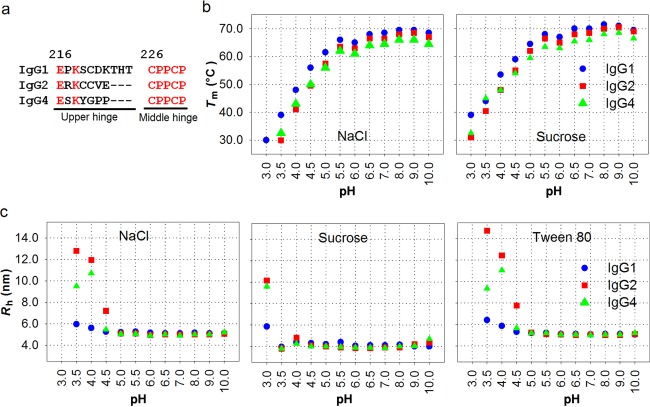
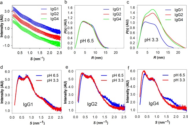
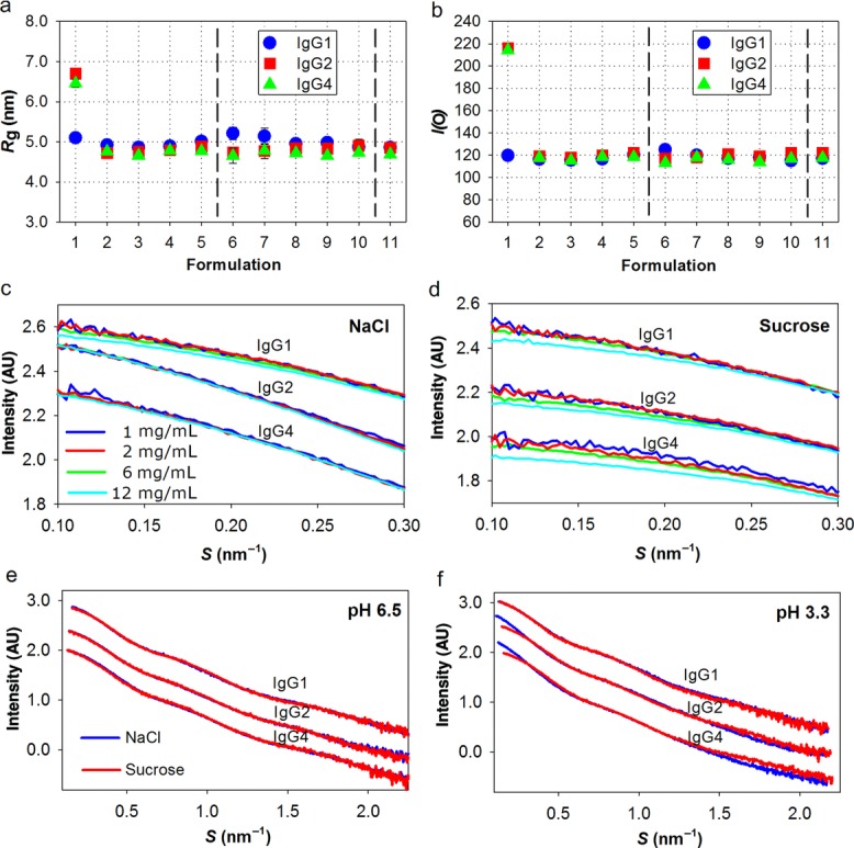
A crucial step in the development of therapeutic monoclonal antibodies is the selection of robust pharmaceutical candidates and screening of efficacious protein formulations to increase the resistance toward physicochemical degradation and aggregation during processing and storage. Here, we introduce small-angle X-ray scattering (SAXS) to characterize antibody solution behavior, which strongly complements conventional biophysical analysis. First, we apply a variety of conventional biophysical techniques for the evaluation of structural, conformational, and colloidal stability and report a systematic comparison between designed humanized IgG1, IgG2, and IgG4 with identical variable regions. Then, the high information content of SAXS data enables sensitive detection of structural differences between three IgG subclasses at neutral pH and rapid formation of dimers of IgG2 and IgG4 at low pH. We reveal subclass-specific variation in intermolecular repulsion already at low and medium protein concentrations, which explains the observed improved stability of IgG1 with respect to aggregation. We show how excipients dramatically influence such repulsive effects, hence demonstrating the potential application of extensive SAXS screening in antibody selection, eventual engineering, and formulation development.
Keywords: IgG antibody; analysis; physicochemical properties; protein aggregation; protein formulation; small-angle X-ray scattering (SAXS); stability.
© 2014 The Authors. Journal of Pharmaceutical Sciences published by Wiley Periodicals, Inc. and the American Pharmacists Association.
Figures
References
-
- Jefferis R. Antibody therapeutics: Isotype and glycoform selection. Expert Opin Biol Ther. 2007;7(9):1401–1413. - PubMed
-
- Chan AC, Carter PJ. Therapeutic antibodies for autoimmunity and inflammation. Nat Rev Immunol. 2010;10(5):301–316. - PubMed
-
- Jiskoot W, Beuvery EC, de Koning AA, Herron JN, Crommelin DJ. Analytical approaches to the study of monoclonal antibody stability. Pharm Res. 1990;7(12):1234–1241. - PubMed
Publication types
MeSH terms
Substances
LinkOut - more resources
Full Text Sources
Other Literature Sources
Molecular Biology Databases
