

Movement-related neuromagnetic fields in preschool age children
- PMID: 24700413
- PMCID: PMC6869527
- DOI: 10.1002/hbm.22518
Movement-related neuromagnetic fields in preschool age children
Abstract
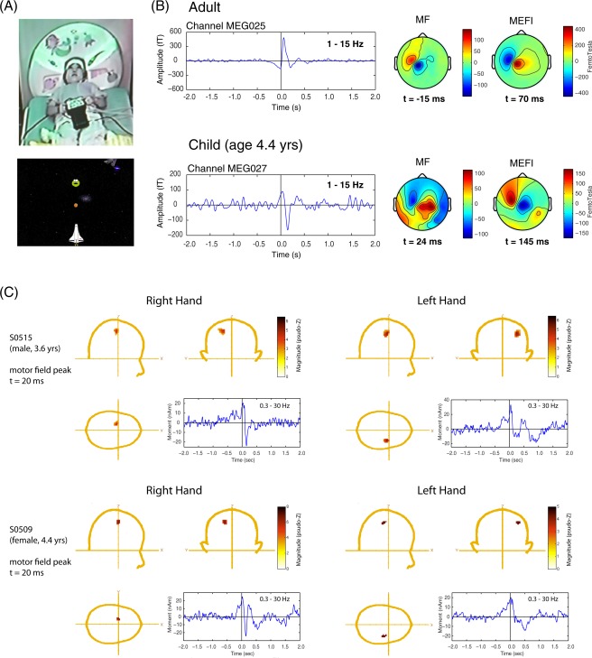
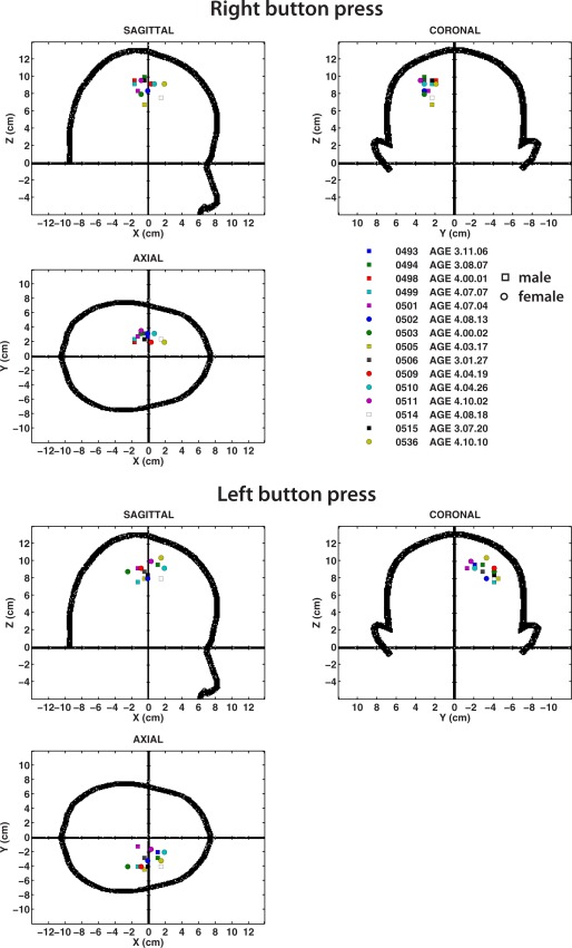
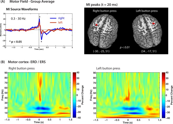
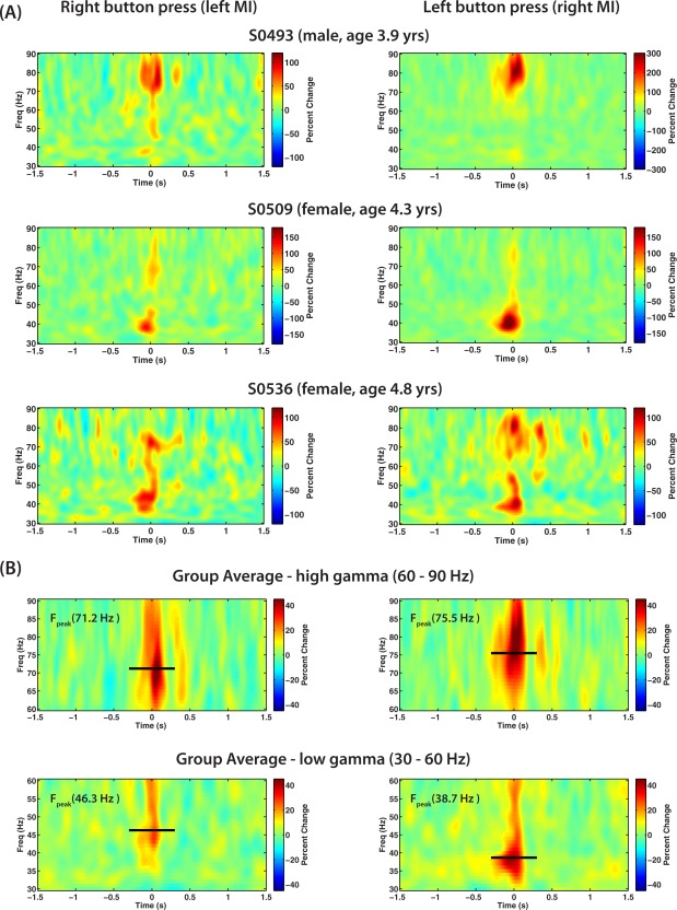
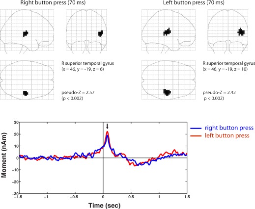
We examined sensorimotor brain activity associated with voluntary movements in preschool children using a customized pediatric magnetoencephalographic system. A videogame-like task was used to generate self-initiated right or left index finger movements in 17 healthy right-handed subjects (8 females, ages 3.2-4.8 years). We successfully identified spatiotemporal patterns of movement-related brain activity in 15/17 children using beamformer source analysis and surrogate MRI spatial normalization. Readiness fields in the contralateral sensorimotor cortex began ∼0.5 s prior to movement onset (motor field, MF), followed by transient movement-evoked fields (MEFs), similar to that observed during self-paced movements in adults, but slightly delayed and with inverted source polarities. We also observed modulation of mu (8-12 Hz) and beta (15-30 Hz) oscillations in sensorimotor cortex with movement, but with different timing and a stronger frequency band coupling compared to that observed in adults. Adult-like high-frequency (70-80 Hz) gamma bursts were detected at movement onset. All children showed activation of the right superior temporal gyrus that was independent of the side of movement, a response that has not been reported in adults. These results provide new insights into the development of movement-related brain function, for an age group in which no previous data exist. The results show that children under 5 years of age have markedly different patterns of movement-related brain activity in comparison to older children and adults, and indicate that significant maturational changes occur in the sensorimotor system between the preschool years and later childhood.
Keywords: MEG; beamformers; development; movement-related fields; mu rhythm; sensorimotor oscillations.
Copyright © 2014 Wiley Periodicals, Inc.
Figures
Similar articles
-
An extended motor network generates beta and gamma oscillatory perturbations during development.Brain Cogn. 2010 Jul;73(2):75-84. doi: 10.1016/j.bandc.2010.03.001. Epub 2010 Apr 24. Brain Cogn. 2010. PMID: 20418003 Free PMC article.
-
Individual differences in motor development during early childhood: An MEG study.Dev Sci. 2020 Sep;23(5):e12935. doi: 10.1111/desc.12935. Epub 2020 Jan 9. Dev Sci. 2020. PMID: 31869490
-
Neuromagnetic Cerebellar Activity Entrains to the Kinematics of Executed Finger Movements.Cerebellum. 2018 Oct;17(5):531-539. doi: 10.1007/s12311-018-0943-4. Cerebellum. 2018. PMID: 29725948
-
Electroencephalographic and magnetoencephalographic studies of motor function.Adv Neurol. 1990;54:193-205. Adv Neurol. 1990. PMID: 2270804 Review.
-
Mu rhythm: State of the art with special focus on cerebral palsy.Ann Phys Rehabil Med. 2020 Oct;63(5):439-446. doi: 10.1016/j.rehab.2019.06.007. Epub 2019 Jul 9. Ann Phys Rehabil Med. 2020. PMID: 31299375 Review.
Cited by
-
Visuomotor Activation of Inhibition-Processing in Pediatric Obsessive Compulsive Disorder: A Magnetoencephalography Study.Front Psychiatry. 2021 Apr 29;12:632736. doi: 10.3389/fpsyt.2021.632736. eCollection 2021. Front Psychiatry. 2021. PMID: 33995145 Free PMC article.
-
The intrahemispheric functional properties of the developing sensorimotor cortex are influenced by maturation.Front Hum Neurosci. 2015 Feb 17;9:39. doi: 10.3389/fnhum.2015.00039. eCollection 2015. Front Hum Neurosci. 2015. PMID: 25741263 Free PMC article.
-
Feasibility and Challenges of Performing Magnetoencephalography Experiments in Children With Arthrogryposis Multiplex Congenita.Front Pediatr. 2021 Oct 4;9:626734. doi: 10.3389/fped.2021.626734. eCollection 2021. Front Pediatr. 2021. PMID: 34671580 Free PMC article.
-
Gamma deficits as a neural signature of cognitive impairment in children treated for brain tumors.J Neurosci. 2014 Jun 25;34(26):8813-24. doi: 10.1523/JNEUROSCI.5220-13.2014. J Neurosci. 2014. PMID: 24966381 Free PMC article.
-
A Novel Candidate Neuromarker of Central Motor Dysfunction in Childhood Apraxia of Speech.J Neurosci. 2025 May 7;45(19):e1471242025. doi: 10.1523/JNEUROSCI.1471-24.2025. J Neurosci. 2025. PMID: 40180575
References
-
- Alegre M, Labarga A, Gurtubay IG, Iriarte J, Malanda A, Artieda J (2002): Beta electroencephalograph changes during passive movements: Sensory afferences contribute to beta event‐related desynchronization in humans. Neurosci Lett 331:29–32. - PubMed
-
- Almli CR, Rivkin MJ, McKinstry RC (2007): The NIH MRI study of normal brain development (Objective‐2): Newborns, infants, toddlers, and preschoolers. NeuroImage 35:308–325. - PubMed
-
- Bardouille T, Picton TW, Ross B (2006): Correlates of eye blinking as determined by synthetic aperture magnetometry. Clin Neurophysiol 117:952–958. - PubMed
Publication types
MeSH terms
LinkOut - more resources
Full Text Sources
Other Literature Sources
Research Materials
